持续性肺部浸润影的病因可以是感染性或非感染性[1]Menendez R, Perpina M, Torres A. Evaluation of non-resolving and progressive pneumonia. Semin Respir Infect. 2003 Jun;18(2):103-11.http://www.ncbi.nlm.nih.gov/pubmed/12840791?tool=bestpractice.com[2]Kuru T, Lynch JP 3rd. Non-resolving or slowly resolving pneumonia. Clin Chest Med. 1999 Sep;20(3):623-51.http://www.ncbi.nlm.nih.gov/pubmed/10516909?tool=bestpractice.com[3]Cunha BA. Slowly resolving and nonresolving pneumonias. Drugs Today (Barc). 2000 Dec;36(12):829-34.http://www.ncbi.nlm.nih.gov/pubmed/12845341?tool=bestpractice.com 临床症状可有或缺如(如咳嗽、咯血)。对于治疗效果不佳,或者胸部影像学无变化甚至进展,或者影像学吸收速度减慢的患者,需对疾病本质进行积极的探寻。[2]Kuru T, Lynch JP 3rd. Non-resolving or slowly resolving pneumonia. Clin Chest Med. 1999 Sep;20(3):623-51.http://www.ncbi.nlm.nih.gov/pubmed/10516909?tool=bestpractice.com
感染性
10%-25%的社区获得性肺炎及30%-60%的医院获得性肺炎的患者表现为不吸收性肺炎,对于治疗反应不佳。[14]Menendez R, Torres A. Treatment failure in community-acquired pneumonia. Chest. 2007 Oct;132(4):1348-55.http://www.ncbi.nlm.nih.gov/pubmed/17934120?tool=bestpractice.com[17]Arancibia F, Ewig S, Martinez JA, et al. Antimicrobial treatment failures in patients with community-acquired pneumonia: causes and prognostic implications. Am J Respir Crit Care Med. 2000 Jul;162(1):154-60.http://www.atsjournals.org/doi/full/10.1164/ajrccm.162.1.9907023http://www.ncbi.nlm.nih.gov/pubmed/10903235?tool=bestpractice.com
社区获得性肺炎可能继发于非典型病原体(例如肺炎支原体、肺炎衣原体、嗜肺军团菌或呼吸道病毒),经常见于在社区附近居住的年轻成人中。
老年和免疫功能抑制患者更容易患上病毒性肺炎,并且叠加其他病原体双重感染(例如金黄色葡萄球菌、流感嗜血杆菌)的情况较为常见。
在疾病流行区域中,如果在常规的抗生素治疗后肺浸润仍持续存在,则应考虑结核病的可能性。真菌性肺炎(例如耶氏肺孢子虫)可能伴有真菌性肉芽肿。病毒性肺炎通常与导致持续性浸润的机化性肺炎有关。其临床表现可能与社区获得性肺炎相似(即流感样疾病,伴有发热、不适、疲乏和咳嗽),影像学上表现为持续性、复发性或游走性、双侧弥漫性肺泡浸润。[18]Gross TJ, Chavis AD, Lynch JP 3rd. Noninfectious pulmonary diseases masquerading as community-acquired pneumonia. Clin Chest Med. 1991 Jun;12(2):363-93.http://www.ncbi.nlm.nih.gov/pubmed/1855377?tool=bestpractice.com[19]Lynch JP 3rd, Sitrin RG. Noninfectious mimics of community acquired pneumonia. Semin Respir Infect. 1993 Mar;8(1):14-45.http://www.ncbi.nlm.nih.gov/pubmed/8372273?tool=bestpractice.com[20]Andrade CR, Ibiapina CC, Champs NS, et al. Avian influenza: the threat of the 21st century. J Bras Pneumol. 2009 May;35(5):470-9.http://www.scielo.br/scielo.php?script=sci_arttext&pid=S1806-37132009000500014&lng=en&nrm=iso&tlng=enhttp://www.ncbi.nlm.nih.gov/pubmed/19547858?tool=bestpractice.com
耐药性或不常见的病原菌(真菌、支原体结核病、诺卡氏菌属、放线菌)可能也会引起持续性肺部浸润。[21]Low DE, Mazzulli T, Marrie T. Progressive and nonresolving pneumonia. Curr Opin Pulm Med. 2005 May;11(3):247-52.http://www.ncbi.nlm.nih.gov/pubmed/15818188?tool=bestpractice.com[22]Weyers CM, Leeper KV. Nonresolving pneumonia. Clin Chest Med. 2005 Mar;26(1):143-58.http://www.ncbi.nlm.nih.gov/pubmed/15802176?tool=bestpractice.com
肺炎的并发症(例如脓胸和肺脓肿)可导致不吸收性肺炎,特别是对于存在共病的患者。计算机体层成像 (CT) 扫描通常在肺泡浸润的基础上、伴有或不伴有支气管充气征皆可诊断。胸腔穿刺术可明确诊断脓胸,不需要其他检查。因肺癌(例如支气管肺癌、支气管类癌、乳头状瘤或者来自乳腺、肾脏或消化道等的转移性病变)导致的阻塞可能会导致复发性梗阻后不吸收性肺炎或脓肿。
 [Figure caption and citation for the preceding image starts]: 不吸收性肺炎的病因由 BMJ Knowledge Centre 根据 Athanasia Pataka 的表格创建 [Citation ends].
免疫抑制性
艾滋病患者 (AIDS) 和非 HIV-免疫功能抑制的宿主可能因多种原因出现持续性肺部浸润(例如,耶氏肺孢子虫性肺炎、结核病)。[23]Boyton RJ. Infectious lung complications in patients with HIV/AIDS. Curr Opin Pulm Med. 2005 May;11(3):203-7.http://www.ncbi.nlm.nih.gov/pubmed/15818180?tool=bestpractice.com[24]Kanmogne GD. Noninfectious pulmonary complications of HIV/AIDS. Curr Opin Pulm Med. 2005 May;11(3):208-12.http://www.ncbi.nlm.nih.gov/pubmed/15818181?tool=bestpractice.com[25]Shorr AF, Susla GM, O'Grady NP. Pulmonary infiltrate in the non-HIV-infected immunocompromised patient: etiologies, diagnostic strategies, and outcomes. Chest. 2004 Jan;125(1):260-71.http://www.ncbi.nlm.nih.gov/pubmed/14718449?tool=bestpractice.com[26]Crawford S. Noninfectious lung disease in the immunocompromised host. Respiration. 1999;66(5):385-95.http://content.karger.com/produktedb/produkte.asp?DOI=29418&typ=pdfhttp://www.ncbi.nlm.nih.gov/pubmed/10516534?tool=bestpractice.com[27]Maschmeyer G, Carratalà J, Buchheidt D, et al. Diagnosis and antimicrobial therapy of lung infiltrates in febrile neutropenic patients (allogeneic SCT excluded): updated guidelines of the Infectious Diseases Working Party (AGIHO) of the German Society of Hematology and Medical Oncology (DGHO). Ann Oncol. 2015 Jan;26(1):21-33.https://www.ncbi.nlm.nih.gov/pmc/articles/PMC4269340/http://www.ncbi.nlm.nih.gov/pubmed/24833776?tool=bestpractice.com[28]Leone S, Nicastri E, Giglio S, et al. Immune reconstitution inflammatory syndrome associated with Mycobacterium tuberculosis infection: a systematic review. Int J Infect Dis. 2010 Apr;14(4):e283-91.http://www.ncbi.nlm.nih.gov/pubmed/19656712?tool=bestpractice.com 卡波西肉瘤可能导致肺部持续性浸润。 [Figure caption and citation for the preceding image starts]: 耶氏肺孢子菌肺炎的后前位胸部 X 线显示出严重的双侧肺间质浸润,伴有囊状影图片由 Matthew Gingo (UPMC) 提供 [Citation ends].
 [Figure caption and citation for the preceding image starts]: 免疫抑制患者持续性肺部浸润影的病因由 BMJ Knowledge Centre 根据 Athanasia Pataka 的表格创建 [Citation ends].
恶性
年龄大于45岁的吸烟者有反复发生的肺炎、咯血和体重减轻需排除恶性肿瘤可能性。[2]Kuru T, Lynch JP 3rd. Non-resolving or slowly resolving pneumonia. Clin Chest Med. 1999 Sep;20(3):623-51.http://www.ncbi.nlm.nih.gov/pubmed/10516909?tool=bestpractice.com 支气管肺泡细胞癌可能表现为伴有支气管充气征的肺泡浸润,类似于不吸收性肺炎。淋巴瘤也可能表现为肺泡浸润,通常伴有支气管充气征。卡波西肉瘤可能会导致肺部持续性浸润,特别对于 AIDS 患者而言。
肺实质性
气道异物可导致阻塞远端反复发生的不吸收性肺炎。[2]Kuru T, Lynch JP 3rd. Non-resolving or slowly resolving pneumonia. Clin Chest Med. 1999 Sep;20(3):623-51.http://www.ncbi.nlm.nih.gov/pubmed/10516909?tool=bestpractice.com[29]Teramoto S, Matsuse T, Ouchi Y. Foreign body aspiration into the lower airways may not be unusual in older adults. Chest. 1998 Jun;113(6):1733-4.http://www.ncbi.nlm.nih.gov/pubmed/9631831?tool=bestpractice.com 吸入性肺炎特征为极度虚弱或瘫痪患者有卧床、口臭和吞咽困难的病史。典型的影像学表现是低垂肺部的浸润影,痰培养为口腔或混合型菌群。
脂质性肺炎由于脂类进入支气管树所致。[30]Spickard A 3rd, Hirschmann JV. Exogenous lipoid pneumonia. Arch Intern Med. 1994 Mar 28;154(6):686-92.http://www.ncbi.nlm.nih.gov/pubmed/8129503?tool=bestpractice.com 脂类可被吸入(如鼻腔滴入油性滴鼻液,偶然吸入化妆油)或填塞阻塞远端的支气管管腔和小气道。 典型的活检病理表现为含有脂泡沫巨噬细胞和巨细胞。 浸润性的淀粉样变性是一种罕见的引起肺部持续浸润影的病因。[31]Utz JP, Swensen SJ, Gertz MA. Pulmonary amyloidosis: the Mayo Clinic experience from 1980 to 1993. Ann Intern Med. 1996 Feb 15;124(4):407-13.http://www.ncbi.nlm.nih.gov/pubmed/8554249?tool=bestpractice.com
心血管系统
心源性肺水肿在COPD患者可表现为不对称的浸润影。[32]Gluecker T, Capasso P, Schnyder P, et al. Clinical and radiologic features of pulmonary edema: a pictorial essay. Radiographics. 1999 Nov-Dec;19(6):1507-31.http://radiographics.rsnajnls.org/cgi/content/full/19/6/1507http://www.ncbi.nlm.nih.gov/pubmed/10555672?tool=bestpractice.com[33]Rudiger A, Gasser S, Fischler M, et al. Comparable increase of B-type natriuretic peptide and amino-terminal pro-B-type natriuretic peptide levels in patients with severe sepsis, septic shock, and acute heart failure. Crit Care Med. 2006 Aug;34(8):2140-4.http://www.ncbi.nlm.nih.gov/pubmed/16763507?tool=bestpractice.com[34]Maisel A. B-type natriuretic peptide levels: diagnostic and prognostic in congestive heart failure: what's next? Circulation. 2002 May 21;105(20):2328-31.http://www.ncbi.nlm.nih.gov/pubmed/12021215?tool=bestpractice.com 如果仅使用抗生素治疗,病变会持续存在。医师通常会在胸部 X 线连续检查 (CXR) 中发现游走性、一过性浸润。仰卧位和俯卧位视图对比可能有助于诊断,因为肺水肿的基底部浸润在俯卧位时通常好转。这些患者通常具有有助于正确诊断的临床体征(例如心脏杂音、S3、外周水肿、颈静脉压升高)。
伴有梗死的肺栓塞可能与延缓吸收性肺炎类似。急性呼吸困难、胸痛和咯血的典型三联征在临床上较为罕见。在血栓栓塞性疾病中,胸部 X 线摄影的检查发现结果可能是非特异性的,伴有空洞性实变影形成和右心扩大。通过常规 X 线摄影或 CT 对胸部进行连续检查显示浸润自周围向中央逐渐消退(冰山融化征)。使用静脉造影剂进行 CT 血管造影或螺旋 CT 扫描可以显示出栓子,可能伴有楔形实变。[35]van Belle A, Buller HR, Huisman MV, et al; Writing Group for the Christopher Study Investigators. Effectiveness of managing suspected pulmonary embolism using an algorithm combining clinical probability, D-dimer testing, and computed tomography. JAMA. 2006 Jan 11;295(2):172-9.http://jama.jamanetwork.com/article.aspx?articleid=202176http://www.ncbi.nlm.nih.gov/pubmed/16403929?tool=bestpractice.com[36]Stein PD, Woodard PK, Weg JG, et al. Diagnostic pathways in acute pulmonary embolism: recommendations of the PIOPED II investigators. Am J Med. 2006 Dec;119(12):1048-55.http://www.ncbi.nlm.nih.gov/pubmed/17145249?tool=bestpractice.com
炎症性/免疫性
结缔组织病可能表现为肺部浸润。系统性红斑狼疮可能累及肺部、肺部脉管系统、胸膜,甚至隔膜。类风湿性关节炎中最常见的肺部表现为间质性肺疾病。皮肌炎或多发性肌炎患者的肺部病变可能是潜在炎症性肌病的并发症亦或是治疗药物的不良反应。硬皮病可能因食道受累而导致反复发作的吸入性肺炎。
间质性肺炎可导致不同的影像学改变,包括持续性浸润影。 这些疾病可以是特发性,也可继发于职业或环境暴露、药物和胶原或血管疾病。[37]Travis WD, Costabel U, Hansell DM, et al; on behalf of the ATS/ERS Committee on Idiopathic Interstitial Pneumonias. An Official American Thoracic Society/European Respiratory Society Statement: Update of the International Multidisciplinary Classification of the Idiopathic Interstitial . Am J Respir Crit Care Med 2013 Sep 15;188(6):733-48.https://www.thoracic.org/statements/resources/interstitial-lung-disease/classification-of-IIPs.pdfhttp://www.ncbi.nlm.nih.gov/pubmed/24032382?tool=bestpractice.com
 [Figure caption and citation for the preceding image starts]: 间质性肺疾病的病因及可能的鉴别诊断由 BMJ Knowledge Centre 根据 Athanasia Pataka 的表格创建 [Citation ends].
机化性肺炎也可能与结缔组织病、其他间质性肺炎、感染、药物或恶性肿瘤相关。其临床表现可能与社区获得性肺炎的表现相似(例如流感样症状,伴有发热、不适、疲乏、咳嗽),在胸部影像学上表现为持续性、反复性或游走性双侧弥漫性肺泡浸润。
弥漫性肺泡出血可由多种疾病引起(如结缔组织血管病、药物、二尖瓣疾病、感染),其胸部影像学可类似于肺炎表现为弥漫性高密度影。[6]Orens JB, Sitrin RG, Lynch JP 3rd. The approach to nonresolving pneumonia. Med Clin North Am. 2004 Feb;52(2):224-9.http://www.ncbi.nlm.nih.gov/pubmed/8078373?tool=bestpractice.com[18]Gross TJ, Chavis AD, Lynch JP 3rd. Noninfectious pulmonary diseases masquerading as community-acquired pneumonia. Clin Chest Med. 1991 Jun;12(2):363-93.http://www.ncbi.nlm.nih.gov/pubmed/1855377?tool=bestpractice.com[19]Lynch JP 3rd, Sitrin RG. Noninfectious mimics of community acquired pneumonia. Semin Respir Infect. 1993 Mar;8(1):14-45.http://www.ncbi.nlm.nih.gov/pubmed/8372273?tool=bestpractice.com[38]Hochberg MC. Updating the American College of Rheumatology revised criteria for the classification of systemic lupus erythematosus. Arthritis Rheum. 1997 Sep;40(9):1725.http://www.ncbi.nlm.nih.gov/pubmed/9324032?tool=bestpractice.com[39]Arnett FC, Edworthy SM, Bloch DA, et al. The American Rheumatism Association 1987 revised criteria for the classification of rheumatoid arthritis. Arthritis Rheum. 1988 Mar;31(3):315-24.http://www.ncbi.nlm.nih.gov/pubmed/3358796?tool=bestpractice.com[40]LeRoy EC, Black C, Fleischmajer R, et al. Scleroderma (systemic sclerosis): classification, subsets, and pathogenesis. J Rheumatol. 1988 Feb;15(2):202-5.http://www.ncbi.nlm.nih.gov/pubmed/3361530?tool=bestpractice.com
系统性血管炎可累及下呼吸道,表现为咳嗽、呼吸困难、咯血、持续性肺泡高密度影、弥漫性磨玻璃影(可能反映肺泡出血)、空洞性结节和胸腔积液。[18]Gross TJ, Chavis AD, Lynch JP 3rd. Noninfectious pulmonary diseases masquerading as community-acquired pneumonia. Clin Chest Med. 1991 Jun;12(2):363-93.http://www.ncbi.nlm.nih.gov/pubmed/1855377?tool=bestpractice.com
肉芽肿性多血管炎(以前被称为韦格纳肉芽肿)是一种主要累及上下呼吸道和肾脏的系统性血管炎。肺部肉芽肿性多血管炎可表现为多灶性肺部受累或孤立性肺部病变,无肺外疾病证据。
结节病可累及任意器官,然而呼吸系统症状伴影像学单纯的肺部浸润影而无特征性的双侧肺门淋巴结肿大,则类似于即使经过抗生素治疗仍持续存在的肺炎。[41]Costabel U. Sarcoidosis: clinical update. Eur Respir J Suppl. 2001 Sep;32:56s-68s.http://erj.ersjournals.com/cgi/content/full/18/32_suppl/56Shttp://www.ncbi.nlm.nih.gov/pubmed/11816825?tool=bestpractice.com
职业和环境暴露史很重要。[42]Infante PF, Newman LS. Beryllium exposure and chronic beryllium disease. Lancet. 2004 Feb 7;363(9407):415-6.http://www.ncbi.nlm.nih.gov/pubmed/14962519?tool=bestpractice.com[43]Begin R, Cantin A, Masse S. Recent advances in the pathogenesis and clinical assessment of mineral dust pneumoconioses: asbestosis, silicosis and coal pneumoconiosis. Eur Respir J. 1989 Nov;2(10):988-1001.http://www.ncbi.nlm.nih.gov/pubmed/2691279?tool=bestpractice.com[44]American Thoracic Society. Diagnosis and initial management of nonmalignant diseases related to asbestos. Am J Respir Crit Care Med. 2004 Sep 15;170(6):691-715.http://www.atsjournals.org/doi/full/10.1164/rccm.200310-1436ST#.U7_nqvldVlwhttp://www.ncbi.nlm.nih.gov/pubmed/15355871?tool=bestpractice.com[45]American Thoracic Society Committee of the Scientific Assembly on Environmental and Occupational Health. Adverse effects of crystalline silica exposure. Am J Respir Crit Care Med. 1997 Feb;155(2):761-8.http://www.ncbi.nlm.nih.gov/pubmed/9032226?tool=bestpractice.com 石棉肺可能导致持续性间质和胸膜浸润,或盘状肺不张(胸膜粘连和纤维化导致肺部畸形和一些小支气管弯曲)。[43]Begin R, Cantin A, Masse S. Recent advances in the pathogenesis and clinical assessment of mineral dust pneumoconioses: asbestosis, silicosis and coal pneumoconiosis. Eur Respir J. 1989 Nov;2(10):988-1001.http://www.ncbi.nlm.nih.gov/pubmed/2691279?tool=bestpractice.com[44]American Thoracic Society. Diagnosis and initial management of nonmalignant diseases related to asbestos. Am J Respir Crit Care Med. 2004 Sep 15;170(6):691-715.http://www.atsjournals.org/doi/full/10.1164/rccm.200310-1436ST#.U7_nqvldVlwhttp://www.ncbi.nlm.nih.gov/pubmed/15355871?tool=bestpractice.com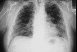 [Figure caption and citation for the preceding image starts]: 后前位胸部 X 线检查表现为双侧基底部线状间质性改变,符合石棉肺表现图片由 Kenneth D. Rosenman (MD) 私人提供 [Citation ends].
胸部 X 线摄影中显示的持续性弥漫性高密度影可能由矽肺所致。[45]American Thoracic Society Committee of the Scientific Assembly on Environmental and Occupational Health. Adverse effects of crystalline silica exposure. Am J Respir Crit Care Med. 1997 Feb;155(2):761-8.http://www.ncbi.nlm.nih.gov/pubmed/9032226?tool=bestpractice.com 进展性的块状纤维化影通常与煤矿工人的尘肺和矽肺有关。
嗜酸粒细胞性肺炎是一组以肺实质内嗜酸性粒细胞异常增多为特征的异质性疾病(伴有或不伴有血清嗜酸粒细胞增多)。它们可能会导致持续性肺部浸润,但大多数经过糖皮质激素治疗后有所改善。[46]Alberts WM. Eosinophilic interstitial lung disease. Curr Opin Pulm Med. 2004 Sep;10(5):419-24.http://www.ncbi.nlm.nih.gov/pubmed/15316442?tool=bestpractice.com[47]Allen JN, Magro CM, King MA. The eosinophilic pneumonias. Semin Respir Crit Care Med. 2002 Apr;23(2):127-34.http://www.ncbi.nlm.nih.gov/pubmed/16088605?tool=bestpractice.com[48]Loffler W. Transient lung infiltrations with blood eosinophilia. Int Arch Allergy Appl Immunol. 1956;8(1-2):54-9.http://www.ncbi.nlm.nih.gov/pubmed/13331628?tool=bestpractice.com[49]Solomon J, Schwarz M. Drug-, toxin-, and radiation therapy-induced eosinophilic pneumonia. Semin Respir Crit Care Med. 2006 Apr;27(2):192-7.http://www.ncbi.nlm.nih.gov/pubmed/16612770?tool=bestpractice.com[50]Zander DS. Allergic bronchopulmonary aspergillosis: an overview. Arch Pathol Lab Med. 2005 Jul;129(7):924-8.http://www.archivesofpathology.org/doi/full/10.1043/1543-2165%282005%29129%5B924%3AABAAO%5D2.0.CO%3B2http://www.ncbi.nlm.nih.gov/pubmed/15974818?tool=bestpractice.com
过敏性支气管肺曲霉病是对定植于支气管的烟曲霉菌的超敏反应。[50]Zander DS. Allergic bronchopulmonary aspergillosis: an overview. Arch Pathol Lab Med. 2005 Jul;129(7):924-8.http://www.archivesofpathology.org/doi/full/10.1043/1543-2165%282005%29129%5B924%3AABAAO%5D2.0.CO%3B2http://www.ncbi.nlm.nih.gov/pubmed/15974818?tool=bestpractice.com 此疾病通常累及哮喘或囊性纤维化患者,亦可见于免疫力下降人群。烟曲霉菌的血清学检查可能有助于诊断。针对此病原体进行的皮肤点刺试验也可作为有用的诊断辅助手段。 [Figure caption and citation for the preceding image starts]: 嗜酸性粒细胞性肺炎患者的胸部 X 线表现图片由 Athanasia Pataka, MD 提供 [Citation ends].
持续暴露于致病抗原的情况下,过敏性肺炎(外源性过敏性肺泡炎)可导致以中上肺区为主的持续性肺部浸润。[51]Lacasse Y, Selman M, Costabel U, et al. Clinical diagnosis of hypersensitivity pneumonitis. Am J Respir Crit Care Med. 2003 Oct 15;168(8):952-8.http://www.atsjournals.org/doi/full/10.1164/rccm.200301-137OChttp://www.ncbi.nlm.nih.gov/pubmed/12842854?tool=bestpractice.com[52]Selman M. Hypersensitivity pneumonitis: a multifaceted deceiving disorder. Clin Chest Med. 2004 Sep;25(3):531-47.http://www.ncbi.nlm.nih.gov/pubmed/15331190?tool=bestpractice.com[53]Silva CI, Churg A, Muller NL. Hypersensitivity pneumonitis: spectrum of high-resolution CT and pathologic findings. AJR Am J Roentgenol. 2007 Feb;188(2):334-44.http://www.ajronline.org/doi/full/10.2214/AJR.05.1826http://www.ncbi.nlm.nih.gov/pubmed/17242239?tool=bestpractice.com
肺泡蛋白沉积症是一种由于表面活性物质清除减少所致的罕见肺部疾病,患者常表现为气短和持续的肺部浸润影。[18]Gross TJ, Chavis AD, Lynch JP 3rd. Noninfectious pulmonary diseases masquerading as community-acquired pneumonia. Clin Chest Med. 1991 Jun;12(2):363-93.http://www.ncbi.nlm.nih.gov/pubmed/1855377?tool=bestpractice.com[54]Shah PL, Hansell D, Lawson PR, et al. Pulmonary alveolar proteinosis: clinical aspects and current concepts on pathogenesis. Thorax. 2000 Jan;55(1):67-77.http://www.pubmedcentral.nih.gov/picrender.fcgi?artid=1745595&blobtype=pdfhttp://www.ncbi.nlm.nih.gov/pubmed/10607805?tool=bestpractice.com[55]Seymour JF, Presneill JJ. Pulmonary alveolar proteinosis: progress in the first 44 years. Am J Respir Crit Care Med. 2002 Jul 15;166(2):215-35.http://www.atsjournals.org/doi/full/10.1164/rccm.2109105http://www.ncbi.nlm.nih.gov/pubmed/12119235?tool=bestpractice.com
浸润性的淀粉样变性是一种罕见的引起肺部持续浸润影的病因。
药源性/医源性
某些药物(如胺碘酮、博来霉素、环磷酰胺、长春新碱、紫杉烷类)和可卡因或其他违禁药品会导致肺部持续浸润影。[19]Lynch JP 3rd, Sitrin RG. Noninfectious mimics of community acquired pneumonia. Semin Respir Infect. 1993 Mar;8(1):14-45.http://www.ncbi.nlm.nih.gov/pubmed/8372273?tool=bestpractice.com[56]Camus P, Bonniaud P, Fanton A, et al. Drug-induced and iatrogenic infiltrative lung disease. Clin Chest Med. 2004 Sep;25(3):479-519.http://www.ncbi.nlm.nih.gov/pubmed/15331188?tool=bestpractice.com[57]Erasmus JJ, McAdams HP, Rossi SE. Drug-induced lung injury. Semin Roentgenol. 2002 Jan;37(1):72-81.http://www.ncbi.nlm.nih.gov/pubmed/11987768?tool=bestpractice.com[58]Camus P. Pneumotox online: the drug-induced respiratory disease website. Vol. 2, 2012 [internet publication].http://www.pneumotox.com正在接受化疗或放疗的恶性肿瘤患者出现持续性肺部浸润既可能是由自身的恶性肿瘤引起,也可能是由治疗的并发症引起。[27]Maschmeyer G, Carratalà J, Buchheidt D, et al. Diagnosis and antimicrobial therapy of lung infiltrates in febrile neutropenic patients (allogeneic SCT excluded): updated guidelines of the Infectious Diseases Working Party (AGIHO) of the German Society of Hematology and Medical Oncology (DGHO). Ann Oncol. 2015 Jan;26(1):21-33.https://www.ncbi.nlm.nih.gov/pmc/articles/PMC4269340/http://www.ncbi.nlm.nih.gov/pubmed/24833776?tool=bestpractice.com[59]Carver JR, Shapiro CL, Ng A, et al. ASCO Cancer Survivorship Expert Panel. American Society of Clinical Oncology clinical evidence review on the ongoing care of adult cancer survivors: cardiac and pulmonary late effects. J Clin Oncol. 2007 Sep 1;25(25):3991-4008.http://jco.ascopubs.org/cgi/content/full/25/25/3991http://www.ncbi.nlm.nih.gov/pubmed/17577017?tool=bestpractice.com [Figure caption and citation for the preceding image starts]: 胺碘酮肺毒性患者的胸部 CT 扫描表现为分布于肺外周的非对称性阴影图片由 Athanasia Pataka, MD 提供 [Citation ends].
 [Figure caption and citation for the preceding image starts]: 胺碘酮肺毒性患者的胸部 X 线表现图片由 Athanasia Pataka, MD 提供 [Citation ends].
 [Figure caption and citation for the preceding image starts]: 药物诱发的肺实质性疾病的可能致病药物由 BMJ Knowledge Centre 根据 Athanasia Pataka 的表格创建 [Citation ends].
