初期表现
多数患者表现很明显,但有一些仅有精神状态改变,而体格检查很少或未发现外伤证据。如果没有可靠的目击者,往往一开始会认为患者是卒中、癫痫、精神病或中毒导致的精神状况改变,最终才会发现隐匿性颅脑外伤。
病史
在初始复苏和治疗的 ABCD 后,对于每一例有脑外伤的患者或原因不明的精神状态改变患者,应有重点的询问病史。关于创伤事件的详细描述应来源于患者本人、家人、急救人员、第一目击者和警察。目击者或熟悉患者的人员有助于详细查明关于创伤事件和环境的详情,也有助于了解患者正常的功能水平。这有助于建立较广泛的鉴别诊断,以防止过早定论的错误。病史应包括以下内容:
体格检查
全身体格检查必须在基本的 ABCD 抢救治疗后进行。除了警惕隐匿性损伤,医生应仔细注意以下体征:
应每 15 分钟进行连续的 GCS 评估和瞳孔检查,直到病情稳定,以确保及时识别神经系统功能恶化。
头颈部手术
检查有无颅神经缺陷、眶周或耳后瘀斑、脑脊液鼻漏或耳漏、鼓室积血(颅底骨折的体征) [Figure caption and citation for the preceding image starts]: 鼓室积血:中耳鼓室内的血液(箭头所示)van Dijk GW. Practical Neurology. 2011;11(1):50-55 [Citation ends].
 [Figure caption and citation for the preceding image starts]: Battle 征:乳突上方的浅表瘀斑van Dijk GW. Practical Neurology. 2011;11(1):50-55 [Citation ends].
视网膜出血的眼底检查(受虐待的体征)[91]Maguire SA, Watts PO, Shaw AD, et al. Retinal haemorrhages and related findings in abusive and non-abusive head trauma: a systematic review. Eye (Lond). 2013 Jan;27(1):28-36.http://www.nature.com/eye/journal/v27/n1/full/eye2012213a.htmlhttp://www.ncbi.nlm.nih.gov/pubmed/23079748?tool=bestpractice.com 和视盘水肿(颅内压升高的体征)
触诊头皮以发现血肿、捻发音、裂伤和骨骼畸形(颅骨骨折的标志)
听诊颈动脉杂音(颈动脉夹层的体征)
评估颈椎压痛、感觉异常、大小便失禁、肢体无力、阴茎异常勃起(脊髓损伤的体征)
在手术室打开硬脑膜并在直视下实施此操作的情况下方可取出明显异物或刺穿物。
需要持续的心功能、血压监测来评估心血管功能。任何低血压事件必须立即处理。[58]Manley G, Knudson MM, Morabito D, et al. Hypotension, hypoxia, and head injury: frequency, duration, and consequences. Arch Surg. 2001 Oct;136(10):1118-23.https://jamanetwork.com/journals/jamasurgery/fullarticle/392263http://www.ncbi.nlm.nih.gov/pubmed/11585502?tool=bestpractice.com[59]Chesnut RM, Marshall LF, Klauber MR, et al. The role of secondary brain injury in determining outcome from severe head injury. J Trauma. 1993 Feb;34(2):216-22.http://www.ncbi.nlm.nih.gov/pubmed/8459458?tool=bestpractice.com
需要连续监测脉搏血氧饱和度和呼吸状态;气管插管的患者,需要连续进行潮气末 CO2 分析。任何缺氧情况必须立即处理。[58]Manley G, Knudson MM, Morabito D, et al. Hypotension, hypoxia, and head injury: frequency, duration, and consequences. Arch Surg. 2001 Oct;136(10):1118-23.https://jamanetwork.com/journals/jamasurgery/fullarticle/392263http://www.ncbi.nlm.nih.gov/pubmed/11585502?tool=bestpractice.com[59]Chesnut RM, Marshall LF, Klauber MR, et al. The role of secondary brain injury in determining outcome from severe head injury. J Trauma. 1993 Feb;34(2):216-22.http://www.ncbi.nlm.nih.gov/pubmed/8459458?tool=bestpractice.com
四肢运动及感觉检查(脊髓损伤的体征)。
GCS 评分和瞳孔检查
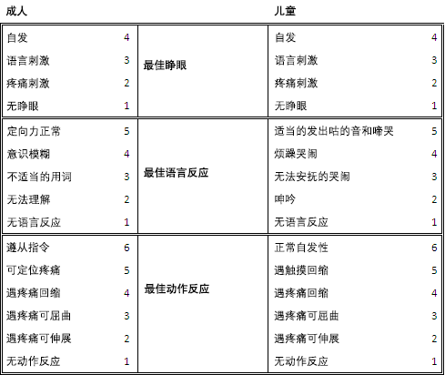
GCS 广泛用于评估 TBI 患者的意识水平,并提供良好的预后信息(当得分很低或很高),使医生能够预期诊断和监测患者的病情。[64]Teasdale G, Jennett B. Assessment of coma and impaired consciousness: a practical scale. Lancet. 1974 Jul 13;2(7872):81-4.http://www.ncbi.nlm.nih.gov/pubmed/4136544?tool=bestpractice.com GCS 13-15 分结局良好,但不能依此排除颅内损伤。评分<9 与临床症状恶化和结局差相关。连续 GCS 检测能够为病情恶化提供临床警示。
GCS 有三个构成要素:最佳睁眼反应 (E)、最佳语言反应 (V) 及最佳运动反应 (M)。每组的评分应分开记录(例如, GCS 10=E3 V4 M3})。运动部分的缺陷与 TBI 患者的不良结局的相关性最大。[92]Hoffmann M, Lefering R, Rueger JM, et al. Pupil evaluation in addition to Glasgow Coma Scale components in prediction of traumatic brain injury and mortality. Br J Surg. 2012 Jan;99(suppl 1):122-30.http://www.ncbi.nlm.nih.gov/pubmed/22441866?tool=bestpractice.com[93]Compagnone C, d'Avella D, Servadei F, et al. Patients with moderate head injury: a prospective multicenter study of 315 patients. Neurosurgery. 2009 Apr;64(4):690-6; discussion 696-7.http://www.ncbi.nlm.nih.gov/pubmed/19197220?tool=bestpractice.com 有口腔、眼外伤的患者或那些进行气管插管、服药的患者或年轻的患者,有时很难评价。但最近的研究表明,乙醇中毒对 GCS 评分的影响不大,除非血液中的乙醇含量>200 mg/dL。[94]Stuke L, Diaz-Arrastia R, Gentilello LM, et al. Effect of alcohol on Glasgow Coma Scale in head-injured patients. Ann Surg. 2007 Apr;245(4):651-5.http://www.ncbi.nlm.nih.gov/pmc/articles/PMC1877033/http://www.ncbi.nlm.nih.gov/pubmed/17414616?tool=bestpractice.com[95]Lange RT, Iverson GL, Brubacher JR, et al. Effect of blood alcohol level on Glasgow Coma Scale scores following traumatic brain injury. Brain Inj. 2010;24(7-8):919-27.http://www.ncbi.nlm.nih.gov/pubmed/20545447?tool=bestpractice.com
 [Figure caption and citation for the preceding image starts]: 成人和小儿 GCS经 Micelle J. Haydel 医生许可使用 [Citation ends].
使用以下评分系统:[45]Taber KH, Warden DL, Hurley RA. Blast-related traumatic brain injury: what is known? J Neuropsychiatry Clin Neurosci. 2006 Spring;18(2):141-5.http://neuro.psychiatryonline.org/article.aspx?articleID=102725http://www.ncbi.nlm.nih.gov/pubmed/16720789?tool=bestpractice.com
GCS 评分 13-15 分提示轻度脑损伤
GCS 评分 9-12 分提示中度脑损伤
GCS 8 分以下提示重度脑损伤
虽然 GCS 13分一直被归为轻度,但许多专家认为应归为中度损伤。值得注意的是,分级的严重性和数字的大小是相反的。医疗团队的不同成员可对患者行连续 GCS 评分以监测神经功能状态;尽管受到质疑,不同人员进行 GCS 评分的可靠性还是很好。[96]Gill MR, Reiley DG, Green SM. Interrater reliability of Glasgow Coma Scale scores in the emergency department. Ann Emerg Med. 2004 Feb;43(2):215-23.http://www.ncbi.nlm.nih.gov/pubmed/14747811?tool=bestpractice.com[97]Tesseris J, Pantazidis N, Routsi C, et al. A comparative study of the Reaction Level Scale (RLS 85) with Glasgow Coma Scale (GCS) and Edinburgh-2 Coma Scale (Modified) (E2CS(M)). Acta Neurochir (Wien). 1991;110(1-2):65-76.http://www.ncbi.nlm.nih.gov/pubmed/1882722?tool=bestpractice.com[98]Elliott M. Interrater reliability of the Glasgow Coma Scale. J Neurosci Nurs. 1996 Aug;28(4):213-4.http://www.ncbi.nlm.nih.gov/pubmed/8880594?tool=bestpractice.com[99]Lindsay KW, Teasdale GM, Knill-Jones RP. Observer variability in assessing the clinical features of subarachnoid hemorrhage. J Neurosurg. 1983 Jan;58(1):57-62.http://www.ncbi.nlm.nih.gov/pubmed/6847910?tool=bestpractice.com[100]Menegazzi J, Davis EA, Sucov AN, et al. Reliability of the Glasgow Coma Scale when used by emergency physicians and paramedics. J Trauma. 1993 Jan;34(1):46-8.http://www.ncbi.nlm.nih.gov/pubmed/8437195?tool=bestpractice.com
瞳孔反射功能作为反映损伤后病理和严重程度的指标,应连续监测。[69]Meyer S, Gibb T, Jurkovich GJ. Evaluation and significance of the pupillary light reflex in trauma patients. Ann Emerg Med. 1993 Jun;22(6):1052-7.http://www.ncbi.nlm.nih.gov/pubmed/8503525?tool=bestpractice.com 瞳孔检查可以在昏迷患者或患者接受神经肌肉阻断剂或镇静剂时评估。[18]Maas AI, Stocchetti N, Bullock R. Moderate and severe traumatic brain injury in adults. Lancet Neurol. 2008 Aug;7(8):728-41.http://www.ncbi.nlm.nih.gov/pubmed/18635021?tool=bestpractice.com 应检查瞳孔大小、对称性、直接/间接对光反射及瞳孔扩大/固定的持续时间。瞳孔反射异常可提示脑疝或脑干损伤。眼眶外伤、药物或直接动眼神经损伤可导致瞳孔变化,但不会出现颅内压增高、脑干病变或脑疝。
在血流动力学稳定无缺氧或低血压的患者中,GCS 评分、瞳孔的评估是最可靠的,这些可能会改变患者的临床检查程序。
实验室检查
基础实验室检查应包括:
全血细胞计数 (FBC) 包括血小板
血清电解质和尿素
血糖
凝血状态:PT、INR、活化 PTT
如果需要,可行血液酒精含量和毒理学筛查
尿液分析。
动脉血气分析在 TBI 不常应用,因为是否需要确保呼吸道通畅,要基于临床发现和预期的住院时间。GCS<8 分的患者,或没有自主呼吸的颅脑外伤患者、不能维持气道的开放或不能维持>90% 的氧饱和度的患者,需要保证气道通畅。
在过去 10 年中,研究者评价了若干种潜在的生物标志物,以鉴定在 CT 显示上有严重颅内损伤的患者。已证实 S-100beta 和胶质纤维酸性蛋白都是具有前景的标志物,但迄今它们的特异性和敏感性均未超过已验证的临床决策指南。[101]Vos PE, Jacobs B, Andriessen TM, et al. GFAP and S100B are biomarkers of traumatic brain injury: an observational cohort study. Neurology. 2010 Nov 16;75(20):1786-93.http://www.ncbi.nlm.nih.gov/pubmed/21079180?tool=bestpractice.com[102]Undén J, Romner B. Can low serum levels of S100B predict normal CT findings after minor head injury in adults?: an evidence-based review and meta-analysis. J Head Trauma Rehabil. 2010 Jul-Aug;25(4):228-40.http://www.ncbi.nlm.nih.gov/pubmed/20611042?tool=bestpractice.com[103]Kotlyar S, Larkin GL, Moore CL, et al. S100b immunoassay: an assessment of diagnostic utility in minor head trauma. J Emerg Med. 2011 Sep;41(3):285-93.http://www.ncbi.nlm.nih.gov/pubmed/20692788?tool=bestpractice.com[104]Schiff L, Hadker N, Weiser S, et al. A literature review of the feasibility of glial fibrillary acidic protein as a biomarker for stroke and traumatic brain injury. Mol Diagn Ther. 2012 Apr 1;16(2):79-92.http://www.ncbi.nlm.nih.gov/pubmed/22497529?tool=bestpractice.com
影像学检查
非增强计算机断层扫描 (CT) 是 TBI 患者首选的影像学检查;它能检测临床上绝大多数重要的损伤,并可以指导 TBI 内科治疗和外科手术的决策。[105]Gentry LR, Godersky JC, Thompson B. Prospective comparative study of intermediate-field MR and CT in the evaluation of closed head trauma. AJR Am J Roentgenol. 1988 Mar;150(3):673-82.http://www.ncbi.nlm.nih.gov/pubmed/3257625?tool=bestpractice.com[106]Moppett IK. Traumatic brain injury: assessment, resuscitation and early management. Br J Anaesth. 2007 Jul;99(1):18-31.http://www.ncbi.nlm.nih.gov/pubmed/17545555?tool=bestpractice.com[107]Vos PE, Alekseenko Y, Battistin L, et al. Mild traumatic brain injury. Eur J Neurol. 2012 Feb;19(2):191-8.http://www.ncbi.nlm.nih.gov/pubmed/22260187?tool=bestpractice.com
美国放射学院 (American College of Radiology) 达成共识,建议继续支持 CT 作为首选检查方法应用于颅脑外伤患者。[108]American College of Radiology. ACR appropriateness criteria: head trauma. 2015 [internet publication].https://acsearch.acr.org/docs/69481/Narrative/ 以下的 CT 表现与颅脑外伤解决较差相关:中线移位、蛛网膜下腔出血进入或压缩/闭塞脑池。[73]Bullock MR, Chesnut R, Ghajar J, et al; Surgical Management of Traumatic Brain Injury Author Group. Surgical management of traumatic parenchymal lesions. Neurosurgery. 2006 Mar;58(3 Suppl):S25-46; discussion Si-iv.http://www.ncbi.nlm.nih.gov/pubmed/16540746?tool=bestpractice.com 当 CT 检查后仍不能明确病变时,可考虑行 MRI 检查,以发现更多的细微病变,例如弥漫性轴索损伤 (DAI)。应立即行 CT 检查的情况包括:所有脑穿透伤患者;怀疑颅底凹陷或开放性骨折;GCS<13 分;或局灶性神经功能缺失的患者。
轻度 TBI 患者 CT 扫描的指南
对于孤立的轻度头部外伤或轻度 TBI 患者,CT 的使用是有争议的,多数的研究已经提出了直接使用 CT 的指导方针。 到目前为止,已出版超过 20 个临床决策规范,[22]Pandor A, Goodacre S, Harnan S, et al. Diagnostic management strategies for adults and children with minor head injury: a systematic review and an economic evaluation. Health Technol Assess. 2011 Aug;15(27):1-202.http://www.journalslibrary.nihr.ac.uk/hta/volume-15/issue-27http://www.ncbi.nlm.nih.gov/pubmed/21806873?tool=bestpractice.com 但新奥尔良标准 (New Orleans Criteria, NOC) 和加拿大头部 CT 规范 (Canadian CT Head Rule, CCHR) 非常出名,因为在存在或不存在意识丧失的 TBI 患者以及 GCS 评分 13-15 分的患者中,进行了多次外部验证,其敏感性高达 99%-100%。[87]Stiell IG, Clement CM, Rowe BH, et al. Comparison of the Canadian CT Head Rule and the New Orleans Criteria in patients with minor head injury. JAMA. 2005 Sep 28;294(12):1511-8.https://jamanetwork.com/journals/jama/fullarticle/201596http://www.ncbi.nlm.nih.gov/pubmed/16189364?tool=bestpractice.com[109]Stein SC, Fabbri A, Servadei F, et al. A critical comparison of clinical decision instruments for computed tomographic scanning in mild closed traumatic brain injury in adolescents and adults. Ann Emerg Med. 2009 Feb;53(2):180-8.http://www.ncbi.nlm.nih.gov/pubmed/18339447?tool=bestpractice.com[110]Smits M, Dippel DW, de Haan GG, et al. External validation of the Canadian CT Head Rule and the New Orleans Criteria for CT scanning in patients with minor head injury. JAMA. 2005 Sep 28;294(12):1519-25.https://jamanetwork.com/journals/jama/fullarticle/201595http://www.ncbi.nlm.nih.gov/pubmed/16189365?tool=bestpractice.com[111]Papa L, Stiell IG, Clement CM, et al. Performance of the Canadian CT Head Rule and the New Orleans Criteria for predicting any traumatic intracranial injury on computed tomography in a United States Level I trauma center. Acad Emerg Med. 2012 Jan;19(1):2-10.http://onlinelibrary.wiley.com/doi/10.1111/j.1553-2712.2011.01247.x/fullhttp://www.ncbi.nlm.nih.gov/pubmed/22251188?tool=bestpractice.com[112]Haydel MJ, Preston CA, Mills TJ, et al. Indications for computed tomography in patients with minor head injury. N Engl J Med. 2000 Jul 13;343(2):100-5.http://www.nejm.org/doi/full/10.1056/NEJM200007133430204#t=articlehttp://www.ncbi.nlm.nih.gov/pubmed/10891517?tool=bestpractice.com 这两项 CT 指南都包括以下变量:某种形式的呕吐、高龄、精神状态改变和体格检查时发现的头部外伤体征。在英国,处理轻微 TBI 患者的 NICE 指南包括来自加拿大头部 CT 规范中的变量。NICE: head injury overview 在美国,对于轻度 TBI 患者的处理,疾病预防控制中心 (Centers for Disease Control and Prevention, CDC) 采用了新奥尔良标准中的变量。CDC: mild TBI pocket guide
加拿大头部 CT 检查规范
对于有轻度颅脑损伤的患者(轻度颅脑损伤患者的定义为 GCS 评分为13-15 分,被目击到有意识丧失、明确的失忆或被目击到定向障碍的患者),如果存在下列任何一种情况,需要进行 CT 检查:[8]Stiell IG, Wells GA, Vandemheen K, et al. The Canadian CT head rule for patients with minor head injury. Lancet. 2001;357(9266):1391-6.http://www.ncbi.nlm.nih.gov/pubmed/11356436?tool=bestpractice.com
高风险(神经系统干预):
中度风险(针对 CT 上显示的脑损伤):
新奥尔良准则
对于存在以下任何一种情况的轻度头部外伤患者(轻度头部损伤的定义为患者到达急诊科时经医师检查,发现简单的神经系统查体结果正常且 GCS 评分为 15 分,但有意识丧失),需进行 CT 检查:[112]Haydel MJ, Preston CA, Mills TJ, et al. Indications for computed tomography in patients with minor head injury. N Engl J Med. 2000 Jul 13;343(2):100-5.http://www.nejm.org/doi/full/10.1056/NEJM200007133430204#t=articlehttp://www.ncbi.nlm.nih.gov/pubmed/10891517?tool=bestpractice.com
高风险(对于神经外科干预而言)
头痛
呕吐
年龄>60 岁
药物或酒精中毒
持续性顺行性遗忘(短期记忆缺失)
锁骨以上创伤性软组织或骨损伤证据
癫痫发作(疑似或可见)。
制定上述标准的过程中还包含了既往的凝血疾病病史作为一种临床参数,但这并未被包括在最后认证中。如果可能,要获取这个疾病史,并考虑进行 CT 扫描。
婴儿和儿童
三项大型的前瞻性研究介绍了用于识别在头部损伤后可获益于 CT 检查儿童的几种临床决策规则。[113]Kuppermann N, Holmes JF, Dayan PS, et al. Identification of children at very low risk of clinically-important brain injuries after head trauma: a prospective cohort study. Lancet. 2009 Oct 3;374(9696):1160-70.http://www.ncbi.nlm.nih.gov/pubmed/19758692?tool=bestpractice.com[114]Osmond MH, Klassen TP, Wells GA, et al. CATCH: a clinical decision rule for the use of computed tomography in children with minor head injury. CMAJ. 2010 Mar 9;182(4):341-8.http://www.cmaj.ca/content/182/4/341.long[115]Dunning J, Daly JP, Lomas JP, et al. Derivation of the children's head injury algorithm for the prediction of important clinical events decision rule for head injury in children. Arch Dis Child. 2006 Nov;91(11):885-91.http://adc.bmj.com/content/91/11/885在 2017 年,一项关于 20,000 名以上儿童的前瞻性研究发现,在识别存在临床重要性 TBI 的方面,儿科急诊应用研究网络 (Pediatric Emergency Care Applied Research Network, PECARN) 临床决策规则具有最高的敏感性。[116]Babl FE, Borland ML, Phillips N, et al. Accuracy of PECARN, CATCH, and CHALICE head injury decision rules in children: a prospective cohort study. Lancet. 2017 Jun 17;389(10087):2393-2402.http://www.sciencedirect.com/science/article/pii/S014067361730555X?via%3Dihub 基于 PECARN 指南,对于 GCS 评分<15 分、精神状态改变(躁动、嗜睡、反复提问、反应迟钝)或明显的颅骨骨折、或怀疑颅底骨折的儿童,均需进行 CT 检查。[113]Kuppermann N, Holmes JF, Dayan PS, et al. Identification of children at very low risk of clinically-important brain injuries after head trauma: a prospective cohort study. Lancet. 2009 Oct 3;374(9696):1160-70.http://www.ncbi.nlm.nih.gov/pubmed/19758692?tool=bestpractice.com其他 CT 检查的指征与年龄有关。
在 PECARN 指南中,对于年龄<2 岁的儿童,其他需行 CT 检查的指征包括:[113]Kuppermann N, Holmes JF, Dayan PS, et al. Identification of children at very low risk of clinically-important brain injuries after head trauma: a prospective cohort study. Lancet. 2009 Oct 3;374(9696):1160-70.http://www.ncbi.nlm.nih.gov/pubmed/19758692?tool=bestpractice.com
在 PECARN 指南中,对于年龄>2 岁的儿童,其他需行 CT 检查的指征包括:[113]Kuppermann N, Holmes JF, Dayan PS, et al. Identification of children at very low risk of clinically-important brain injuries after head trauma: a prospective cohort study. Lancet. 2009 Oct 3;374(9696):1160-70.http://www.ncbi.nlm.nih.gov/pubmed/19758692?tool=bestpractice.com
轻度外伤性颅脑损伤(脑震荡)
轻度外伤性颅脑损伤的诊断 (mTBI) 依赖于详细的病史和检查。影像学未发现异常的情况下,不能排除 TBI 诊断。患者的病史和家属的询问是在形成诊断的重要部分。[117]Ruff RM, Iverson GL, Barth JT, et al; NAN Policy and Planning Committee. Recommendations for diagnosing a mild traumatic brain injury: a National Academy of Neuropsychology education paper. Arch Clin Neuropsychol. 2009 Feb;24(1):3-10.http://www.ncbi.nlm.nih.gov/pubmed/19395352?tool=bestpractice.com 根据 TBI 的定义,应对意识丧失、逆行性遗忘症、创伤后健忘症、意识不清和定向力障碍,和局灶性神经功能缺损进行仔细评估。[117]Ruff RM, Iverson GL, Barth JT, et al; NAN Policy and Planning Committee. Recommendations for diagnosing a mild traumatic brain injury: a National Academy of Neuropsychology education paper. Arch Clin Neuropsychol. 2009 Feb;24(1):3-10.http://www.ncbi.nlm.nih.gov/pubmed/19395352?tool=bestpractice.com 此外,症状和体征可能是由酒精或药物的影响引起。[117]Ruff RM, Iverson GL, Barth JT, et al; NAN Policy and Planning Committee. Recommendations for diagnosing a mild traumatic brain injury: a National Academy of Neuropsychology education paper. Arch Clin Neuropsychol. 2009 Feb;24(1):3-10.http://www.ncbi.nlm.nih.gov/pubmed/19395352?tool=bestpractice.com 目前,诊断轻度 TBI 主要根据临床病史和体格检查。轻度颅脑外伤后,CT 检查结果通常是正常的,但相当数量的患者遗留神经认知功能障碍,神经科医师的随访和考虑进行弥散张量成像可能使患者受益。[118]Kraus MF, Susmaras T, Caughlin BP, et al. White matter integrity and cognition in chronic traumatic brain injury: a diffusion tensor imaging study. Brain. 2007 Oct;130(Pt 10):2508-19.https://academic.oup.com/brain/article-lookup/doi/10.1093/brain/awm216http://www.ncbi.nlm.nih.gov/pubmed/17872928?tool=bestpractice.com[119]Aoki Y, Inokuchi R, Gunshin M, et al. Diffusion tensor imaging studies of mild traumatic brain injury: a meta-analysis. J Neurol Neurosurg Psychiatry. 2012 Sep;83(9):870-6.http://jnnp.bmj.com/content/83/9/870.longhttp://www.ncbi.nlm.nih.gov/pubmed/22797288?tool=bestpractice.com
 [Figure caption and citation for the preceding image starts]: 鼓室积血:中耳鼓室内的血液(箭头所示)van Dijk GW. Practical Neurology. 2011;11(1):50-55 [Citation ends].
 [Figure caption and citation for the preceding image starts]: Battle 征:乳突上方的浅表瘀斑van Dijk GW. Practical Neurology. 2011;11(1):50-55 [Citation ends].
 [Figure caption and citation for the preceding image starts]: 成人和小儿 GCS经 Micelle J. Haydel 医生许可使用 [Citation ends].