孤立性肺结节是一种常见的胸部 X 线检查发现,随着胸部计算机体层成像 (CT) 的普遍使用,这一类型的结节越来越多见。[6]Klein JS, Braff S. Imaging evaluation of the solitary pulmonary nodule. Clin Chest Med. 2008 Mar;29(1):15-38, v.http://www.ncbi.nlm.nih.gov/pubmed/18267182?tool=bestpractice.com 临床医生的初始目标是区分肺结节的良恶性。 如高度怀疑结节为恶性,大多数病例需尽快行切除术。
临床标准和影像学发现被用于判断结节恶性的可能性并作出诊疗决定。最常见的鉴别诊断包括原发性肺癌、转移性肺癌、感染性和炎症性疾病以及血管炎,如肉芽肿性多血管炎(以前被称为韦格纳肉芽肿)。
恶性肺结节的临床特征
以下三个临床特征是恶性肺结节的独立预测指标:
1. 高龄
孤立性肺结节的恶性概率与患者的年龄直接相关。 可通过计算似然比来量化年龄对孤立性肺结节恶性概率的影响。 似然比等于真阳性率(敏感性)除于假阳性率(1-特异性)。 似然比高有助于诊断某种疾病,似然比低有助于排除某种疾病,而似然比为1时对判断恶性概率无价值。[7]Deeks JJ, Altman DG. Diagnostic tests 4: likelihood ratios. BMJ. 2004 Jul 17;329(7458):168-9.http://www.ncbi.nlm.nih.gov/pubmed/15258077?tool=bestpractice.com 现已计算出年龄<30 岁人群的似然比为 0.05,而年龄>70 岁人群的似然比为 4.16-5.7。[8]Gurney JW. Determining the likelihood of malignancy in solitary pulmonary nodules with Bayesian analysis. Part I. Theory. Radiology. 1993 Feb;186(2):405-13.http://www.ncbi.nlm.nih.gov/pubmed/8421743?tool=bestpractice.com[9]Cummings SR, Lillington GA, Richard RJ. Estimating the probability of malignancy in solitary pulmonary nodules: a Bayesian approach. Am Rev Respir Dis. 1986 Sep;134(3):449-52.http://www.ncbi.nlm.nih.gov/pubmed/3752700?tool=bestpractice.com 多变量分析已证实,年龄是恶性孤立性肺结节的独立危险因素。[10]Swensen SJ, Silverstein MD, Ilstrup DM, et al. The probability of malignancy in solitary pulmonary nodules: application to small radiologically indeterminate nodules. Arch Intern Med. 1997 Apr 28;157(8):849-55.http://www.ncbi.nlm.nih.gov/pubmed/9129544?tool=bestpractice.com
2. 吸烟状况
吸烟一直被认为是肺癌的危险因素,它也是恶性孤立性肺结节的一个独立预测指标。[10]Swensen SJ, Silverstein MD, Ilstrup DM, et al. The probability of malignancy in solitary pulmonary nodules: application to small radiologically indeterminate nodules. Arch Intern Med. 1997 Apr 28;157(8):849-55.http://www.ncbi.nlm.nih.gov/pubmed/9129544?tool=bestpractice.com 在从不吸烟的人群中,一个恶性孤立性肺结节的似然比为0.15-0.19。[8]Gurney JW. Determining the likelihood of malignancy in solitary pulmonary nodules with Bayesian analysis. Part I. Theory. Radiology. 1993 Feb;186(2):405-13.http://www.ncbi.nlm.nih.gov/pubmed/8421743?tool=bestpractice.com[9]Cummings SR, Lillington GA, Richard RJ. Estimating the probability of malignancy in solitary pulmonary nodules: a Bayesian approach. Am Rev Respir Dis. 1986 Sep;134(3):449-52.http://www.ncbi.nlm.nih.gov/pubmed/3752700?tool=bestpractice.com 似然比随着吸烟量的增加而增大(例如,每天吸烟 10-20 支者的似然比为 1.0,而每天吸烟> 40 支者的似然比为 3.9)且随着戒烟时间的延长而减小(例如戒烟<3 年者的似然比为 1.4,而戒烟>13 年以上者的似然比为 0.1)。
还有证据表明吸烟是肺癌的独立危险因素。虽然对于吸烟<5 支/天的吸烟者,罹患肺癌的相对危险度为 1.57(置信区间:0.67-3.66,无统计学显著性),但对于更大量吸烟者(>5 支/天),罹患肺癌的相对危险度增至 3.24(置信区间:1.01-10.4)。[11]Iribarren C, Tekawa IS, Sidney S, et al. Effect of cigar smoking on the risk of cardiovascular disease, chronic obstructive pulmonary disease, and cancer in men. N Engl J Med. 1999 Jun 10;340(23):1773-80.http://www.nejm.org/doi/full/10.1056/NEJM199906103402301http://www.ncbi.nlm.nih.gov/pubmed/10362820?tool=bestpractice.com 在吸烟者中,一个孤立性肺结节的恶性似然比范围为 0.3-1.0。这意味着对于一个孤立性肺结节为恶性的可能性,吸烟者是非吸烟患者的 2 到 5 倍(非吸烟者似然比为 0.15-0.19)。[8]Gurney JW. Determining the likelihood of malignancy in solitary pulmonary nodules with Bayesian analysis. Part I. Theory. Radiology. 1993 Feb;186(2):405-13.http://www.ncbi.nlm.nih.gov/pubmed/8421743?tool=bestpractice.com[9]Cummings SR, Lillington GA, Richard RJ. Estimating the probability of malignancy in solitary pulmonary nodules: a Bayesian approach. Am Rev Respir Dis. 1986 Sep;134(3):449-52.http://www.ncbi.nlm.nih.gov/pubmed/3752700?tool=bestpractice.com 因此,与从不吸烟者相比,吸烟可使罹患肺癌的风险增加 2 到 5 倍,但是仅考虑计算似然比并不足以判定孤立性肺结节的性质。
3. 罹患其他恶性肿瘤
既往有恶性肿瘤病史(>5年以前)是恶性孤立性肺结节的独立危险因素。[10]Swensen SJ, Silverstein MD, Ilstrup DM, et al. The probability of malignancy in solitary pulmonary nodules: application to small radiologically indeterminate nodules. Arch Intern Med. 1997 Apr 28;157(8):849-55.http://www.ncbi.nlm.nih.gov/pubmed/9129544?tool=bestpractice.com 有恶性肿瘤病史的患者,随先前恶性肿瘤的性质而异,孤立性肺结节为恶性的似然比为3.82~4.95。[8]Gurney JW. Determining the likelihood of malignancy in solitary pulmonary nodules with Bayesian analysis. Part I. Theory. Radiology. 1993 Feb;186(2):405-13.http://www.ncbi.nlm.nih.gov/pubmed/8421743?tool=bestpractice.com[10]Swensen SJ, Silverstein MD, Ilstrup DM, et al. The probability of malignancy in solitary pulmonary nodules: application to small radiologically indeterminate nodules. Arch Intern Med. 1997 Apr 28;157(8):849-55.http://www.ncbi.nlm.nih.gov/pubmed/9129544?tool=bestpractice.com
评估孤立性肺结节时,还应考虑肺癌的其他已知危险因素,例如存在中度或重度的阻塞性肺病。[12]Mannino DM, Aguayo SM, Petty TL, et al. Low lung function and incident lung cancer in the United States: data from the first National Health and Nutrition Examination Survey follow-up. Arch Intern Med. 2003 Jun 23;163(12):1475-80.http://archinte.jamanetwork.com/article.aspx?articleid=215738http://www.ncbi.nlm.nih.gov/pubmed/12824098?tool=bestpractice.com 以及细小颗粒物暴露或与硫氧化物相关的污染暴露,[13]Pope CA 3rd, Burnett RT, Thun MJ, et al. Lung cancer, cardiopulmonary mortality, and long-term exposure to fine particulate air pollution. JAMA. 2002 Mar 6;287(9):1132-41.http://jama.jamanetwork.com/article.aspx?articleid=194704http://www.ncbi.nlm.nih.gov/pubmed/11879110?tool=bestpractice.com 这些可以作为孤立性肺结节恶性的预测指标,但尚无关于这方面的研究。其他病史特征(例如咯血)已被用于临床预测模式,但尚未被证实是恶性孤立性肺结节的独立预测指标。年龄小于 30 岁的终生不吸烟者是唯一能使临床医生作出随访观察而无需进一步检查决定的临床特征组合。所有其他临床特征被用于计算癌症的可能性,以作出进一步决定。
恶性孤立性肺结节的影像学特征
数个影像学标准被用于估算孤立性肺结节的恶性概率。
钙化类型
一种中央高密度的,分层的,软骨样钙化类型(常称为爆米花样钙化)或一种弥散性的钙化类型强烈提示为良性结节。 良性钙化类型诊断恶性肺结节的似然比接近零。[8]Gurney JW. Determining the likelihood of malignancy in solitary pulmonary nodules with Bayesian analysis. Part I. Theory. Radiology. 1993 Feb;186(2):405-13.http://www.ncbi.nlm.nih.gov/pubmed/8421743?tool=bestpractice.com 即使如此,约 13% 的恶性肺结节为非良性钙化类型(见 E 和 F)。 [Figure caption and citation for the preceding image starts]: A-D:良性结节的钙化类型;E、F:可能见于恶性结节Mazzone P.J., Stoller J.K. Semin Thorac Cardiovasc Surg. 2002;14:250-260; 经许可后使用 [Citation ends].
生长速率
生长非常缓慢的孤立性肺结节(体积倍增时间大于500天)或,与之相反,生长非常快速的孤立性肺结节(体积倍增时间小于30天)提示为良性病变,虽然这并非是绝对的法则。 传统观点认为两年内稳定的肺结节可认为是良性病变仅适用于实性孤立性肺结节。 然而此观点已受到质疑且不适用于磨玻璃结节。[14]Yankelevitz DF, Henschke CI. Does 2-year stability imply that pulmonary nodules are benign? AJR Am J Roentgenol. 1997 Feb;168(2):325-8.http://www.ajronline.org/doi/pdf/10.2214/ajr.168.2.9016198http://www.ncbi.nlm.nih.gov/pubmed/9016198?tool=bestpractice.com 此外,临床医生应该记住,在 X 线胸片上球形病灶直径增加 30% 意味着体积增大一倍。
边缘特征
大小
总的说来,良性病变的大小要小于恶性病变。 例如,直径小于2cm的结节仅20%为恶性结节,而80%的直径大于3cm的肺肿块为恶性病变。 然而,很多孤立性肺小结节为恶性病变,故不能仅凭病灶大小区分良恶性。[15]Davis EW, Katz S, Peabody JW Jr. The solitary pulmonary nodule: a ten-year study based on 215 cases. J Thorac Surg. 1956 Dec;32(6):728-70.http://www.ncbi.nlm.nih.gov/pubmed/13377448?tool=bestpractice.com[16]Steele JD. The solitary pulmonary nodule: report of a cooperative study of resected asymptomatic solitary pulmonary nodules in males. J Thorac Cardiovasc Surg. 1963 Jul;46:21-39.http://www.ncbi.nlm.nih.gov/pubmed/14044162?tool=bestpractice.com
部位
上叶和中叶孤立性肺结节恶性的似然比为 1.2-1.6。[8]Gurney JW. Determining the likelihood of malignancy in solitary pulmonary nodules with Bayesian analysis. Part I. Theory. Radiology. 1993 Feb;186(2):405-13.http://www.ncbi.nlm.nih.gov/pubmed/8421743?tool=bestpractice.com[16]Steele JD. The solitary pulmonary nodule: report of a cooperative study of resected asymptomatic solitary pulmonary nodules in males. J Thorac Cardiovasc Surg. 1963 Jul;46:21-39.http://www.ncbi.nlm.nih.gov/pubmed/14044162?tool=bestpractice.com 病灶位于上叶是恶性肺结节的独立危险因素。[10]Swensen SJ, Silverstein MD, Ilstrup DM, et al. The probability of malignancy in solitary pulmonary nodules: application to small radiologically indeterminate nodules. Arch Intern Med. 1997 Apr 28;157(8):849-55.http://www.ncbi.nlm.nih.gov/pubmed/9129544?tool=bestpractice.com
诊断策略
针对评估孤立性肺结节,适当的诊断性检查包括:[17]Gould MK, Donington J, Lynch WR, et al. Evaluation of individuals with pulmonary nodules: when is it lung cancer? Diagnosis and management of lung cancer, 3rd ed: American College of Chest Physicians evidence-based clinical practice guidelines. Chest. 2013 May;143(5 Suppl):e93S-e120S.http://www.ncbi.nlm.nih.gov/pubmed/23649456?tool=bestpractice.com
与之前的胸部 X 线检查资料或计算机体层成像 (CT) 扫描结果进行比较。如果结节以前就存在并在两年内无变化,除非高度怀疑为生长缓慢的恶性肿瘤(例如类癌、细支气管肺泡癌),否则可进行观察且无需进一步检查。传统观点认为两年内稳定的肺结节可被认为是良性病变,仅适用于实性孤立性肺结节。然而此观点已受到质疑,且应谨慎用于有磨玻璃样阴影的患者中。[14]Yankelevitz DF, Henschke CI. Does 2-year stability imply that pulmonary nodules are benign? AJR Am J Roentgenol. 1997 Feb;168(2):325-8.http://www.ajronline.org/doi/pdf/10.2214/ajr.168.2.9016198http://www.ncbi.nlm.nih.gov/pubmed/9016198?tool=bestpractice.com
如果通过胸部 X 线检查发现病灶,则应进行胸部 CT 检查。高分辨 CT 扫描能更准确地发现结节的特征,例如大小、边缘、钙化和位置。它还有助于识别其他结节,并可能有助于对恶性肿瘤进行分期。
评估恶性肿瘤的临床概率。根据患者的病史特征(如年龄、吸烟史、合并其他恶性肿瘤)和影像学特征(如钙化、生长速率、边缘和大小),临床医生可预估恶性概率,进而根据患者的意愿和整体健康状况,与患者讨论对观察以及无创或有创检查的需求。
确定恶性概率(验前概率)
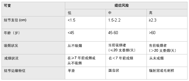
根据临床和影像学特征计算恶性病变的验前概率有助于临床医生判定哪些患者可以安全地随访(如恶性概率小于10%),哪些患者需要手术切除(如恶性概率大于90%),剩余的患者为不确定的(如恶性概率大于10%并小于90%)。 重要的是需记住概率阈值因临床医生和患者的情况而异,且需考虑相关的并发症。 不幸的是,这种决策方法并不准确。 基于Bayesian技术[9]Cummings SR, Lillington GA, Richard RJ. Estimating the probability of malignancy in solitary pulmonary nodules: a Bayesian approach. Am Rev Respir Dis. 1986 Sep;134(3):449-52.http://www.ncbi.nlm.nih.gov/pubmed/3752700?tool=bestpractice.com 或多变量逻辑回归分析[10]Swensen SJ, Silverstein MD, Ilstrup DM, et al. The probability of malignancy in solitary pulmonary nodules: application to small radiologically indeterminate nodules. Arch Intern Med. 1997 Apr 28;157(8):849-55.http://www.ncbi.nlm.nih.gov/pubmed/9129544?tool=bestpractice.com 已显示出一定前景,但在预测恶性肿瘤时并不优于专家级别的临床医生。[18]Swensen SJ, Silverstein MD, Edell ES, et. al. Solitary pulmonary nodules: clinical prediction model versus physicians. Mayo Clin Proc. 1999;74:319-29.http://www.ncbi.nlm.nih.gov/pubmed/10221459?tool=bestpractice.com 可能有助于计算癌症验前概率的两种方法如下。
临床决策  [Figure caption and citation for the preceding image starts]: 恶性孤立性肺结节的验前概率Ost D., Fein A.M., Feinsilver S.H.N Engl J Med.2003;348:2535-2542 [Citation ends].
贝叶斯定理或多元回归方程。多元回归方程基于患者年龄、吸烟史、肺外恶性肿瘤史、结节的直径、结节的边缘特征和在肺部的位置,计算孤立性肺结节为恶性的验前概率。[10]Swensen SJ, Silverstein MD, Ilstrup DM, et al. The probability of malignancy in solitary pulmonary nodules: application to small radiologically indeterminate nodules. Arch Intern Med. 1997 Apr 28;157(8):849-55.http://www.ncbi.nlm.nih.gov/pubmed/9129544?tool=bestpractice.com 计算公式如下:恶性概率=e^x/(1 + e^x)。
其中:
连续 CT 扫描
如果恶性概率低(如基于患者的临床特征几率小于10%),则可行CT扫描随访。 分别在3个月、6个月、12个月和24个月时随访CT是监测结节生长的标准做法。 随访两年是基于生长非常快速的孤立性肺结节(体积倍增时间小于30天)或生长非常缓慢的孤立性肺结节(体积倍增时间大于500天)提示为良性病变。 一项随访10年的研究发现,恶性肿瘤的倍增时间通常为60到150天,平均值为120天。[19]Higgins GA, Shields TW, Keehn RJ. The solitary pulmonary nodule: ten-year follow-up of Veterans Administration-Armed Forces Cooperative Study. Arch Surg. 1975 May;110(5):570-5.http://www.ncbi.nlm.nih.gov/pubmed/1093512?tool=bestpractice.com 确切的倍增时间因发现的不同组织学细胞类型而异,小细胞肺癌为 30 天,而腺癌为 161 天。[20]Geddes DM. The natural history of lung cancer: a review based on rates of tumour growth. Br J Dis Chest. 1979 Jan;73(1):1-17.http://www.ncbi.nlm.nih.gov/pubmed/435370?tool=bestpractice.com 基于生长速率,如果 <7 天观察到有生长或≥ 465 天仍未观察到生长,则恶性病变的似然比接近零。[8]Gurney JW. Determining the likelihood of malignancy in solitary pulmonary nodules with Bayesian analysis. Part I. Theory. Radiology. 1993 Feb;186(2):405-13.http://www.ncbi.nlm.nih.gov/pubmed/8421743?tool=bestpractice.com 因此,病灶生长速度很快或两年内稳定不变是得到认可的良性病变指征。由于磨玻璃样阴影生长十分缓慢,故该期限并不适用。
一项大型多中心研究表明当使用15HU(Hounsfield unit)作为界点的增强CT扫描诊断恶性病变的敏感性为98%,特异性为58%。 这些结果是鼓舞人心的,还需进一步研究明确增强CT在诊断程序中的作用。[21]American College of Radiology. ACR appropriateness criteria: radiographically detected solitary pulmonary nodule. 2012 [internet publication].https://acsearch.acr.org/docs/69455/Narrative/
CT 扫描时偶然发现的小结节 (<1 cm)
对于偶然发现的肺部微小结节(直径< 1 cm)需用不同的方法来评估。因不相关病因(例如呼吸困难、胸痛、腹痛)进行 CT 影像学检查时发现肺结节的情况越来越多见。对于年龄>35 岁、无临床症状、无恶性肿瘤病史、CT 偶然发现直径<1 cm 的肺结节患者,Fleischner 学会 (Fleischner Society) 建议根据患者的结节大小和危险因素决定 CT 扫描随访情况。[22]MacMahon H, Naidich DP, Goo JM, et al. Guidelines for management of incidental pulmonary nodules detected on CT images: from the Fleischner Society 2017. Radiology. 2017 Jul;284(1):228-43.https://pubs.rsna.org/doi/10.1148/radiol.2017161659http://www.ncbi.nlm.nih.gov/pubmed/28240562?tool=bestpractice.com
对于考虑为低风险的患者,2017 年 Fleischner 学会指南推荐:[22]MacMahon H, Naidich DP, Goo JM, et al. Guidelines for management of incidental pulmonary nodules detected on CT images: from the Fleischner Society 2017. Radiology. 2017 Jul;284(1):228-43.https://pubs.rsna.org/doi/10.1148/radiol.2017161659http://www.ncbi.nlm.nih.gov/pubmed/28240562?tool=bestpractice.com
对于被考虑为高风险的患者,2017 年的 Fleischner 学会指南推荐:[22]MacMahon H, Naidich DP, Goo JM, et al. Guidelines for management of incidental pulmonary nodules detected on CT images: from the Fleischner Society 2017. Radiology. 2017 Jul;284(1):228-43.https://pubs.rsna.org/doi/10.1148/radiol.2017161659http://www.ncbi.nlm.nih.gov/pubmed/28240562?tool=bestpractice.com
一项大型、多中心、随机对照临床试验表明用低剂量 CT 进行筛查可降低肺癌的死亡率。该试验入组了 53,454 例肺癌风险增加的患者,并证实死亡率相对降低 20%。[23]Aberle DR, Adams AM, Berg CD, et al; National Lung Screening Trial Research Team. Reduced lung-cancer mortality with low-dose computed tomographic screening. N Engl J Med. 2011 Aug 4;365(5):395-409.http://www.nejm.org/doi/full/10.1056/NEJMoa1102873http://www.ncbi.nlm.nih.gov/pubmed/21714641?tool=bestpractice.com 然而,还存在很多疑问,包括关于这些发现的正确实施、随访时长及这种筛查项目的成本效益。
一项研究描述了预测计算器对估算吸烟者和既往吸烟者接受低剂量 CT 筛查发现的肺结节恶性风险的能力。[24]McWilliams A, Tammemagi MC, Mayo JR, et al. Probability of cancer in pulmonary nodules detected on first screening CT. N Engl J Med. 2013 Sep 5;369(10):910-9.http://www.nejm.org/doi/full/10.1056/NEJMoa1214726#t=articlehttp://www.ncbi.nlm.nih.gov/pubmed/24004118?tool=bestpractice.com 恶性肺结节的预测因素包括:年龄、女性、肺癌家族史、肺气肿、大小、位于肺上叶、部分实性结节、结节计数更低和毛刺征。正如预期,所有受试者均为吸烟者或曾经吸烟者;因此,吸烟史并不作为预测模型的一部分。
18-氟脱氧葡萄糖-PET成像
18-氟脱氧葡萄糖 (FDG) PET 扫描是发现肿瘤的有效方法。氟脱氧葡萄糖在糖酵解时被细胞摄取,不能进入糖酵解途径。在高代谢率的细胞中可见摄取增多,例如肿瘤和炎症部位。报告的敏感性范围为 83%-100%,发现肿瘤的特异性在 50%-100% 之间不等。一项 meta 分析显示敏感性为 96.8% 而特异性为 77.8%。[25]Gould MK, Maclean CC, Kuschner WG, et al. Accuracy of positron emission tomography for diagnosis of pulmonary nodules and mass lesions: a meta-analysis. JAMA. 2001 Feb 21;285(7):914-24.http://jama.jamanetwork.com/article.aspx?articleid=193567http://www.ncbi.nlm.nih.gov/pubmed/11180735?tool=bestpractice.com 诊断恶性病变的阳性似然比为4.36而阴性似然比为0.04。 不幸的是代谢活跃的良性结节可出现假阳性结果(如感染)而代谢相对较慢的肿瘤如细支气管肺泡细胞癌或小肿瘤(5-8mm)可出现假阴性结果。[26]Lowe VJ, Naunheim KS. Current role of positron emission tomography in thoracic oncology. Thorax. 1998 Aug;53(8):703-12.http://www.ncbi.nlm.nih.gov/pubmed/9828860?tool=bestpractice.com PET 扫描还对肿瘤分期有用,且在排除纵隔受累方面有非常高的阴性预测值。[27]Pieterman RM, van Putten JW, Meuzelaar JJ, et al. Preoperative staging of non-small cell lung cancer with positron-emission tomography. N Engl J Med. 2000 Jul 27;343(4):254-61.http://www.nejm.org/doi/full/10.1056/NEJM200007273430404http://www.ncbi.nlm.nih.gov/pubmed/10911007?tool=bestpractice.com [Figure caption and citation for the preceding image starts]: PET-CT显示右肺下叶支气管周围结节高摄取18-FDG(疑为恶性病变);随后经电磁导航支气管镜检查证实为鳞癌由Erik E. Folch, MD提供 [Citation ends].
有创性检查
采取的方法取决于结节的大小和位置、当地的流行病学以及可用的人力和物力资源,因此存在差异。胸内科医生的建议十分宝贵。
两种最常用的方法是:
纤维支气管镜检查。 可通过灌洗、刷检、经支气管肺活检和经支气管针吸活检取得标本。 成功率取决于结节的位置和大小,有无支气管直接进入病灶(支气管征),以及当地应用支气管镜的技术和能力。 该技术的敏感性变化较大,据报道对于2~3cm的结节的敏感性为40%到70%。[28]Swensen SJ, Jett JR, Payne WS, et al. An integrated approach to evaluation of the solitary pulmonary nodule. Mayo Clin Proc. 1990 Feb;65(2):173-86.http://www.ncbi.nlm.nih.gov/pubmed/2248630?tool=bestpractice.com[29]Schreiber G, McCrory DC. Performance characteristics of different modalities for diagnosis of suspected lung cancer: summary of published evidence. Chest. 2003 Jan;123(suppl 1):115S-128S.http://www.ncbi.nlm.nih.gov/pubmed/12527571?tool=bestpractice.com[30]Hergott CA, Tremblay A. Role of bronchoscopy in the evaluation of solitary pulmonary nodules. Clin Chest Med. 2010 Mar;31(1):49-63.http://www.ncbi.nlm.nih.gov/pubmed/20172432?tool=bestpractice.com 先进的支气管镜技术(例如超细支气管镜、电磁导航支气管镜、支气管内超声)可及性已变得更为广泛。这些技术在肺结节评估中的应用,取决于每项技术的可用性以及是否有经过培训的临床医生。支气管镜检查的主要优势在于,与其他采集标本的方法相比,其发生并发症的概率极低。一项meta分析显示引导下支气管镜技术总的诊断率为70%,并随着病灶大小的增加而增加。 39项研究3052例肺结节的气胸总发生率为1.5%。 这项研究表明引导下或先进的支气管镜技术在诊断肺结节上优于传统支气管镜。[31]Wang Memoli JS, Nietert PJ, Silvestri GA. Meta-analysis of guided bronchoscopy for the evaluation of the pulmonary nodule. Chest. 2012 Aug;142(2):385-93.http://journal.publications.chestnet.org/article.aspx?articleid=1262330http://www.ncbi.nlm.nih.gov/pubmed/21980059?tool=bestpractice.com
经胸壁穿刺针吸活检(Transthoracic needle aspiration,TTNA)。 CT引导下TTNA有助于诊断不明确的孤立性肺结节性质。 敏感性为72%到99%,平均值为90%。[29]Schreiber G, McCrory DC. Performance characteristics of different modalities for diagnosis of suspected lung cancer: summary of published evidence. Chest. 2003 Jan;123(suppl 1):115S-128S.http://www.ncbi.nlm.nih.gov/pubmed/12527571?tool=bestpractice.com 然而由于该方法取得的标本量较少故较难诊断良性病变。[32]Ost D, Fein A. Evaluation and management of the solitary pulmonary nodule. Am J Respir Crit Care Med. 2000 Sep;162(3 Pt 1):782-7.http://www.atsjournals.org/doi/full/10.1164/ajrccm.162.3.9812152http://www.ncbi.nlm.nih.gov/pubmed/10988081?tool=bestpractice.com TTNA 最常见的并发症是气胸,发生率为 20% 到 30%。
理论上,应在胸内科医生及放射科医生会诊后决定采用何种检查方法。一般而言,由于与 TTNA 相关的气胸相对发生率和其他技术难题,故通常首选经支气管镜肺活检。TTNA 推荐用于经支气管镜肺活检阴性但仍需进一步明确诊断的病例。需结合当地的技术条件及考虑每一次操作对患者可能的获益和风险来作决定。
先进的支气管镜技提高了软式纤维支气管镜的诊断率,包括:
电磁导航支气管镜。 利用CT行气道重建并借助特殊的软件进行计算可在支气管镜检查过程中判断与靶病灶的距离。 患者置身于一个低场强的电磁定位板上,从而围绕胸腔产生一个电磁场。 位于延长工作槽远端的位置检测器评估电磁场内的方向和位置。 一旦位置检测器探头到达靶目标,固定延长工作槽,活检装置如活检钳、细针、刷子可通过延长工作槽到达靶病灶。 这项技术将纤维支气管镜的诊断率提高至74%。[33]Gildea TR, Mazzone PJ, Karnak D, et al. Electromagnetic navigation diagnostic bronchoscopy: a prospective study. Am J Respir Crit Care Med. 2006 Nov 1;174(9):982-9.http://www.atsjournals.org/doi/full/10.1164/rccm.200603-344OChttp://www.ncbi.nlm.nih.gov/pubmed/16873767?tool=bestpractice.com
外周支气管内超声 (peripheral endobronchial ultrasound, pEBUS) 引导下软式纤维支气管镜检查。支气管内超声使得操作者可看到软式纤维支气管镜所达远端部位的远端结构。在仔细解读胸部 CT 结果后,将一个直径为 1.4 mm 或 1.8 mm 的 20 mHz 探头置于引导鞘内引入感兴趣的胸部节段。一旦到达指定目标区域,探头即从引导鞘中移除。此时,可引入活检钳、细针、刷子进行取样。该技术的总体诊断率为 34% 到 84%。[30]Hergott CA, Tremblay A. Role of bronchoscopy in the evaluation of solitary pulmonary nodules. Clin Chest Med. 2010 Mar;31(1):49-63.http://www.ncbi.nlm.nih.gov/pubmed/20172432?tool=bestpractice.com[34]Herth FJ, Ernst A, Becker HD. Endobronchial ultrasound-guided transbronchial lung biopsy in solitary pulmonary nodules and peripheral lesions. Eur Respir J. 2002 Oct;20(4):972-4.http://erj.ersjournals.com/content/20/4/972.longhttp://www.ncbi.nlm.nih.gov/pubmed/12412691?tool=bestpractice.com
电视胸腔镜手术或开胸手术切除
经上述检查方法仍未能明确诊断的孤立性肺结节病例可选择电视胸腔镜手术(Video-assisted thoracoscopic surgery,VATS)或开胸手术以诊断及治疗。 VATS比开胸术相对安全。 VATS的并发症较少但受限于结节的位置。 如果结节不靠近胸膜则不易通过VATS切除。[35]Bernard A. Resection of pulmonary nodules using video-assisted thoracic surgery. Ann Thorac Surg. 1996 Jan;61(1):202-4.http://www.ncbi.nlm.nih.gov/pubmed/8561553?tool=bestpractice.com
讨论患者的手术选择时需认识到肺叶切除术的死亡率为3%到7%,而楔形切除或结节切除术的死亡率为0.5%到1%。[2]Tan BB, Flaherty KR, Kazerooni EA, et al. The solitary pulmonary nodule. Chest. 2003 Jan;123(1 Suppl):89S-96S.http://www.ncbi.nlm.nih.gov/pubmed/12527568?tool=bestpractice.com[16]Steele JD. The solitary pulmonary nodule: report of a cooperative study of resected asymptomatic solitary pulmonary nodules in males. J Thorac Cardiovasc Surg. 1963 Jul;46:21-39.http://www.ncbi.nlm.nih.gov/pubmed/14044162?tool=bestpractice.com[36]Landreneau RJ, Hazelrigg SR, Ferson PF, et al. Thoracoscopic resection of 85 pulmonary lesions. Ann Thorac Surg. 1992 Sep;54(3):415-9.http://www.ncbi.nlm.nih.gov/pubmed/1510507?tool=bestpractice.com 当外科手术活检明确孤立性肺结节为非小细胞肺癌时,需行肺叶切除术和系统性纵隔淋巴结清扫术。需谨记外科手术前需进行心肺功能的评估。
诊断法则
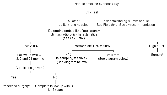
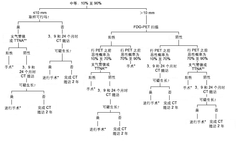
孤立性肺结节的诊断和治疗策略十分复杂。
 [Figure caption and citation for the preceding image starts]: 处理孤立性肺结节的方法。*如果心肺功能允许且临床分期提示局限性病变由Erik E. Folch和Peter J. Mazzone创建 [Citation ends].
 [Figure caption and citation for the preceding image starts]: 如何处理孤立性肺结节。 *如果心肺功能允许且临床分期为局限性病变。 **取样方法的选择基于结节的大小、位置及当地的技术水平并需与胸外科医生和/或介入放射科医生进行磋商。 FDG,18-氟脱氧葡萄糖;TTNA,经胸壁穿刺针吸活检由Erik E. Folch和Peter J. Mazzone创建 [Citation ends].
没有一种方法适用于所有患者。[37]Stern RG. The incidental solitary pulmonary nodule: algorithms, options, and patient choice. Am J Med. 2012 Mar;125(3):221-2.http://www.ncbi.nlm.nih.gov/pubmed/22340914?tool=bestpractice.com 我们推荐根据临床和影像学特征,从估算恶性概率开始采用妥善的处理方法。需基于检查的可及性、当地活检技术水平、合并症及患者意愿来选择额外的检查方法或治疗。[17]Gould MK, Donington J, Lynch WR, et al. Evaluation of individuals with pulmonary nodules: when is it lung cancer? Diagnosis and management of lung cancer, 3rd ed: American College of Chest Physicians evidence-based clinical practice guidelines. Chest. 2013 May;143(5 Suppl):e93S-e120S.http://www.ncbi.nlm.nih.gov/pubmed/23649456?tool=bestpractice.com
诊断流程基于“所有高度可疑的恶性孤立性肺结节都应尽可能地被切除”这一前提。针对最有可能为良性的结节,应进行观察以及临床和影像学随访。针对余下的或性质待定的孤立性肺结节,需由临床医生判断该结节不是恶性。
 [Figure caption and citation for the preceding image starts]: A-D:良性结节的钙化类型;E、F:可能见于恶性结节Mazzone P.J., Stoller J.K. Semin Thorac Cardiovasc Surg. 2002;14:250-260; 经许可后使用 [Citation ends].
 [Figure caption and citation for the preceding image starts]: 计算机体层成像 (CT) 扫描显示右上肺叶毛刺样孤立性肺结节(疑为恶性);随后经支气管镜活检证实为鳞状细胞癌由Erik E. Folch, MD提供 [Citation ends].
 [Figure caption and citation for the preceding image starts]: 恶性孤立性肺结节的验前概率Ost D., Fein A.M., Feinsilver S.H.N Engl J Med.2003;348:2535-2542 [Citation ends].
 [Figure caption and citation for the preceding image starts]: PET-CT显示右肺下叶支气管周围结节高摄取18-FDG(疑为恶性病变);随后经电磁导航支气管镜检查证实为鳞癌由Erik E. Folch, MD提供 [Citation ends].
 [Figure caption and citation for the preceding image starts]: 处理孤立性肺结节的方法。*如果心肺功能允许且临床分期提示局限性病变由Erik E. Folch和Peter J. Mazzone创建 [Citation ends].
 [Figure caption and citation for the preceding image starts]: 如何处理孤立性肺结节。 *如果心肺功能允许且临床分期为局限性病变。 **取样方法的选择基于结节的大小、位置及当地的技术水平并需与胸外科医生和/或介入放射科医生进行磋商。 FDG,18-氟脱氧葡萄糖;TTNA,经胸壁穿刺针吸活检由Erik E. Folch和Peter J. Mazzone创建 [Citation ends].