由于症状表现多样且病程具有不确定性,诊断、分类和治疗该病颇具挑战性。 对苔藓样糠疹的综合性诊断方法是组织学检查。 组织学检查也可以鉴别苔藓样糠疹的不同亚型。[4]Bowers S, Warshaw EM. Pityriasis lichenoides and its subtypes. J Am Acad Dermatol. 2006 Oct;55(4):557-72;http://www.ncbi.nlm.nih.gov/pubmed/17010734?tool=bestpractice.com 皮肤镜检查可鉴别慢性苔藓样糠疹和滴状银屑病,减少了需要活组织检查的病例数。[15]Errichetti E, Lacarrubba F, Micali G et al. Differentiation of pityriasis lichenoides chronica from guttate psoriasis by dermoscopy. Clin Exp Dermatol. 2015 Oct;40(7):804-6.http://www.ncbi.nlm.nih.gov/pubmed/25682853?tool=bestpractice.com
如果怀疑病因是感染,建议检测抗链球菌溶血素 O 滴度、咽拭子培养、EBV 抗体、嗜异凝集抗体或嗜异性抗体、Sabin-Feldman 弓形虫染色,并进行弓形体病、肝炎和 HIV 的血清学诊断。[4]Bowers S, Warshaw EM. Pityriasis lichenoides and its subtypes. J Am Acad Dermatol. 2006 Oct;55(4):557-72;http://www.ncbi.nlm.nih.gov/pubmed/17010734?tool=bestpractice.com
临床评估
急性苔藓痘疮样糠疹(亦称 Mucha-Habermann 病)表现为大量的红斑样小丘疹,迅速发展为不同阶段的各种形态的皮损,如水疱、脓疱、出血性硬皮丘疹和浅溃疡。[3]Blyumin ML, Khachemoune A. What caused these excoriated papules? Diagnosis: pityriasis lichenoides et varioliformis acuta (PLEVA). Skin and Aging. 2004;12:99-102. 皮损易发于躯干前侧、弯侧表面和四肢近端,但也可能出现在其他部位。皮损可能伴有烧灼感、瘙痒和轻度全身症状,但极为少见。[1]Khachemoune A, Blyumin ML. Pityriasis lichenoides: pathophysiology, classification, and treatment review. Am J Clin Dermatol. 2007;8(1):29-36.http://www.ncbi.nlm.nih.gov/pubmed/17298104?tool=bestpractice.com 低热、淋巴结肿大、不适感、肌痛、关节痛和头痛可能先于皮疹发生,或与皮疹同时发生,表明有潜在的传染源。
慢性苔藓样糠疹表现为逐渐出现的粉红色至棕色斑疹和丘疹,中心附着薄的鳞屑。[3]Blyumin ML, Khachemoune A. What caused these excoriated papules? Diagnosis: pityriasis lichenoides et varioliformis acuta (PLEVA). Skin and Aging. 2004;12:99-102.大部分皮损通常散在分布于躯干和四肢近端。
发热溃疡坏死性糠疹是一种极为罕见的、严重的苔藓样糠疹,表现为突然出现的全身性皮疹,呈紫黑色,可见溃疡坏死和结痂,可能累及黏膜。[3]Blyumin ML, Khachemoune A. What caused these excoriated papules? Diagnosis: pityriasis lichenoides et varioliformis acuta (PLEVA). Skin and Aging. 2004;12:99-102.[16]Cozzio A, Hafner J, Kempf W, et al. Febrile ulceronecrotic Mucha-Habermann disease with clonality: A cutaneous T-cell lymphoma entity? J Am Acad Dermatol. 2004 Dec;51(6):1014-7.http://www.ncbi.nlm.nih.gov/pubmed/15583604?tool=bestpractice.com 还有皮损疼痛、瘙痒、发热、全身症状和多器官损伤等其他明显表现。[1]Khachemoune A, Blyumin ML. Pityriasis lichenoides: pathophysiology, classification, and treatment review. Am J Clin Dermatol. 2007;8(1):29-36.http://www.ncbi.nlm.nih.gov/pubmed/17298104?tool=bestpractice.com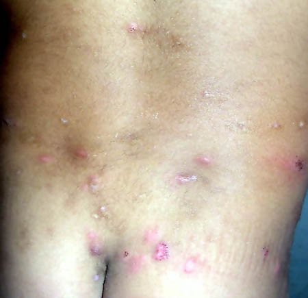 [Figure caption and citation for the preceding image starts]: 散布于儿童背部和臀部的丘疹,颜色呈粉红色至红色,可见坏死、剥脱和结痂,为急性苔藓痘疮样糠疹的表现。摘自纽约州立大学州南部医学中心(布鲁克林) A. Khachemoune 博士收集的资料。 [Citation ends].
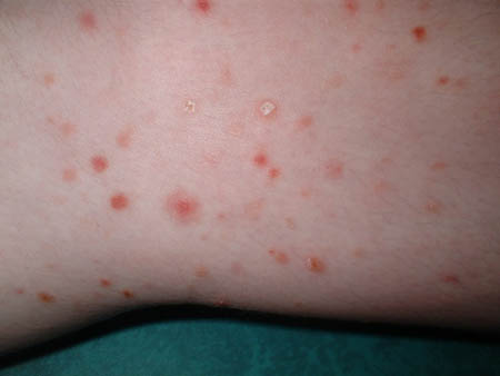 [Figure caption and citation for the preceding image starts]: 慢性苔藓样糠疹近照(云母状鳞屑)摘自纽约州立大学州南部医学中心(布鲁克林) A. Khachemoune 博士收集的资料。 [Citation ends].
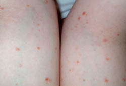 [Figure caption and citation for the preceding image starts]: 慢性苔藓样糠疹,儿童手臂上出现粉红色至浅棕色的扁平小丘疹摘自纽约州立大学州南部医学中心(布鲁克林) A. Khachemoune 博士收集的资料。 [Citation ends].
危险因素
苔藓样糠疹多发于童年晚期和成年早期,且男性易感性稍高。 HIV 患者患苔藓样糠疹的易感性也稍高。
组织病理学检查
组织病理学检查可确认苔藓样糠疹的诊断。 急性苔藓痘疮样糠疹的特点为:弥散性的以 CD8+ 为主的真皮淋巴细胞浸润,浸润呈楔形,较为致密,主要集中在基底层,通过淋巴细胞的胞吐作用进入表皮。[17]Nair PS. A clinical and histopathological study of pityriasis lichenoides. Indian J Dermatol Venereol Leprol. 2007 Mar-Apr;73(2):100-2.http://www.ijdvl.com/article.asp?issn=0378-6323;year=2007;volume=73;issue=2;spage=100;epage=102;aulast=Nairhttp://www.ncbi.nlm.nih.gov/pubmed/17456915?tool=bestpractice.com 表皮上可见凋亡和坏死的角质细胞、嗜中性粒细胞包涵体、海绵状结构和局灶性角化不全。慢性苔藓样糠疹的组织病理学检查显示,在真皮浅层血管周围可见以 CD4+ T 细胞为主的带状淋巴细胞浸润,较为稀疏。在表皮层也可见轻度角化不全的鳞屑和极轻微的角质层坏死和海绵状结构。发热溃疡坏死性糠疹表现为更为显著的淋巴细胞和中性粒细胞浸润,明显的表皮坏死和溃疡,以及白细胞破碎性血管炎。[16]Cozzio A, Hafner J, Kempf W, et al. Febrile ulceronecrotic Mucha-Habermann disease with clonality: A cutaneous T-cell lymphoma entity? J Am Acad Dermatol. 2004 Dec;51(6):1014-7.http://www.ncbi.nlm.nih.gov/pubmed/15583604?tool=bestpractice.com[17]Nair PS. A clinical and histopathological study of pityriasis lichenoides. Indian J Dermatol Venereol Leprol. 2007 Mar-Apr;73(2):100-2.http://www.ijdvl.com/article.asp?issn=0378-6323;year=2007;volume=73;issue=2;spage=100;epage=102;aulast=Nairhttp://www.ncbi.nlm.nih.gov/pubmed/17456915?tool=bestpractice.com
血清学检查
如果怀疑是由传染性病原体诱发的,可以进行相关病原体的血清学检测,以帮助查明病因。 发热溃疡坏死性 Mucha-Habermann 病可以表现出多种实验室检查异常,如白细胞增多、低白蛋白血症、红细胞沉降率 (ESR)、C 反应蛋白 (CRP) 和乳酸脱氢酶升高,这些都是全身性炎症的标志,并有助于确定诊断。[1]Khachemoune A, Blyumin ML. Pityriasis lichenoides: pathophysiology, classification, and treatment review. Am J Clin Dermatol. 2007;8(1):29-36.http://www.ncbi.nlm.nih.gov/pubmed/17298104?tool=bestpractice.com[18]Kim HS, Yu DS, Kim JW. A case of febrile ulceronecrotic Mucha-Habermann's disease successfully treated with oral cyclosporine. JEADV. 2007 Feb;21(2):272-3.http://www.ncbi.nlm.nih.gov/pubmed/17243979?tool=bestpractice.com
