疾病进展迅速使得早期识别尤为重要。当蜂窝织炎患者出现包括全身炎症反应综合征在内的全身性症状时,应考虑发生坏死性筋膜炎的可能性,其中全身炎症反应综合征 (systemic inflammatory response syndrome, SIRS) 的定义为出现以下≥2 种情况:体温>38°C 或<36°C,心率>90 次/分,呼吸频率>20 次/分,或者白细胞计数 [WBC]>12×10⁹/L 或<4×10⁹/L 或未成熟中性粒细胞 [杆状核细胞] 占比>10%)。发热、心动过速、呼吸急促和低血压是至关重要的异常生命体征,其出现可能提示蜂窝织炎患者可能存在需要迅速开展手术评估的广泛性坏死性筋膜炎。蜂窝织炎患者可能存在坏死性筋膜炎的其他症状包括头晕目眩、心悸、恶心、呕吐或谵妄。
病史
询问近期有无皮肤损伤或破溃、创伤、手术、静脉使用药物、水痘或带状疱疹。注意这种诱发性损伤可能是轻微的(例如,昆虫咬伤),也可能未被患者提起。[4]Pasternack MS, Swartz MN. Cellulitis, necrotizing fasciitis, and subcutaneous tissue infections. In: Bennett JE, Dolin R, Blaser MJ, eds. Mandell, Douglas, and Bennett’s principles and practice of infectious diseases. Philadelphia, PA: Elsevier; 2015:1194-215.[12]Stevens DL, Aldape MJ, Bryant AE. Necrotizing fasciitis, gas gangrene, myositis and myonecrosis. In: Cohen J, Powderly WG, Opal SM, eds. Infectious diseases. Amsterdam, Netherlands: Elsevier; 2017:95–103.e1. 暴露史可能偶尔会有所帮助(例如:与嗜水气单胞菌相关的淡水暴露、盐水暴露或与创伤弧菌相关的进食生牡蛎);但经验性的抗生素选择应该是广谱的,而不应仅仅受限于既往暴露因素的提示。[12]Stevens DL, Aldape MJ, Bryant AE. Necrotizing fasciitis, gas gangrene, myositis and myonecrosis. In: Cohen J, Powderly WG, Opal SM, eds. Infectious diseases. Amsterdam, Netherlands: Elsevier; 2017:95–103.e1.[18]Kuo YL, Shieh SJ, Chiu HY, et al. Necrotizing fasciitis caused by Vibrio vulnificus: epidemiology, clinical findings, treatment and prevention. Eur J Clin Microbiol Infect Dis. 2007 Nov;26(11):785-92.
体格检查
蜂窝织炎部位的麻木或剧痛也可能提示潜在的皮下感染。[2]Childers BJ, Potyondy LD, Nachreiner R, et al. Necrotizing fasciitis: a fourteen-year retrospective study of 163 consecutive patients. Am Surg. 2002 Feb;68(2):109-16.[3]Hasham S, Matteucci P, Stanley PR, et al. Necrotising fasciitis. BMJ. 2005 Apr 9;330(7495):830-3. [Erratum in: BMJ. 2005 May 14;330(7500):1143.][4]Pasternack MS, Swartz MN. Cellulitis, necrotizing fasciitis, and subcutaneous tissue infections. In: Bennett JE, Dolin R, Blaser MJ, eds. Mandell, Douglas, and Bennett’s principles and practice of infectious diseases. Philadelphia, PA: Elsevier; 2015:1194-215.[5]Stevens DL, Bisno AL, Chambers HF, et al. Practice guidelines for the diagnosis and management of skin and soft tissue infections: 2014 update by the Infectious Diseases Society of America. Clin Infect Dis. 2014 Jul 15;59(2):e10-52.https://academic.oup.com/cid/article/59/2/e10/2895845[6]Bisno AL, Stevens DL. Streptococcal infections of skin and soft tissues. N Engl J Med. 1996 Jan 25;334(4):240-5.[19]Cheung JP, Fung B, Tang WM, et al. A review of necrotising fasciitis in the extremities. Hong Kong Med J. 2009 Feb;15(1):44-52.http://www.hkmj.org/system/files/hkm0902p44.pdf[20]Angoules AG, Kontakis G, Drakoulakis E, et al. Necrotising fasciitis of upper and lower limb: a systematic review. Injury. 2007 Dec;38 Suppl 5:S19-26.[21]Endorf FW, Cancio LC, Klein MB. Necrotizing soft-tissue infections: clinical guidelines. J Burn Care Res. 2009 Sep-Oct;30(5):769-75. 坏死性筋膜炎时出现的疼痛可能与可见的皮肤变化不成比例。应该注意的是,坏死性筋膜炎患者的表面皮肤可能表现正常,A 族链球菌所致坏死性筋膜炎的表面皮肤变化是一种晚期体征。对于某些病例,对蜂窝织炎区域进行皮肤检查可能发现捻发音、水疱、大疱、皮肤变为灰白色或水肿。诸如液体漏出和水肿等细微的皮肤变化发生于大疱和发红等明显皮肤变化之前。
大约有一半的筋膜炎病例发生于四肢,其余的集中在会阴部、躯干和头颈部。[2]Childers BJ, Potyondy LD, Nachreiner R, et al. Necrotizing fasciitis: a fourteen-year retrospective study of 163 consecutive patients. Am Surg. 2002 Feb;68(2):109-16.[3]Hasham S, Matteucci P, Stanley PR, et al. Necrotising fasciitis. BMJ. 2005 Apr 9;330(7495):830-3. [Erratum in: BMJ. 2005 May 14;330(7500):1143.][4]Pasternack MS, Swartz MN. Cellulitis, necrotizing fasciitis, and subcutaneous tissue infections. In: Bennett JE, Dolin R, Blaser MJ, eds. Mandell, Douglas, and Bennett’s principles and practice of infectious diseases. Philadelphia, PA: Elsevier; 2015:1194-215.[5]Stevens DL, Bisno AL, Chambers HF, et al. Practice guidelines for the diagnosis and management of skin and soft tissue infections: 2014 update by the Infectious Diseases Society of America. Clin Infect Dis. 2014 Jul 15;59(2):e10-52.https://academic.oup.com/cid/article/59/2/e10/2895845[6]Bisno AL, Stevens DL. Streptococcal infections of skin and soft tissues. N Engl J Med. 1996 Jan 25;334(4):240-5.[19]Cheung JP, Fung B, Tang WM, et al. A review of necrotising fasciitis in the extremities. Hong Kong Med J. 2009 Feb;15(1):44-52.http://www.hkmj.org/system/files/hkm0902p44.pdf[20]Angoules AG, Kontakis G, Drakoulakis E, et al. Necrotising fasciitis of upper and lower limb: a systematic review. Injury. 2007 Dec;38 Suppl 5:S19-26. A 族链球菌所致坏死性筋膜炎的最常见部位是大腿,发生于四肢特别是手臂的坏死性筋膜炎更可能由 A 族链球菌所致,而非由多重细菌感染所致。近期有腹部手术史,或腹股沟部位的坏死性筋膜炎最可能由多重细菌感染导致。部分坏死性筋膜炎病例可能因为感染持续扩散而发生相关肌炎。与多重细菌感染相比,这种情况更常见于 A 族链球菌感染。
实验室评估和影像学评估
对所有疑似坏死性筋膜炎的入院患者,均应检查全血细胞计数和白细胞分类计数、尿素、电解质、肌酐和 C 反应蛋白 (C-reactive protein, CRP) 浓度。还应该获得所有患者的动脉血气分析结果,以评估呼吸状态;血培养有助于识别致病病原体。
坏死性筋膜炎通常伴有一系列非特异性实验室检查结果的异常,包括:
若临床适当,理想情况下应对所有疑似坏死性筋膜炎患者进行 X 线平片检查或 CT/MRI(如果可用)。然而,如果无法立即行影像学检查,可能不建议推迟手术,例如当坏死性筋膜炎的风险较高以及患者临床病情不稳定时。[5]Stevens DL, Bisno AL, Chambers HF, et al. Practice guidelines for the diagnosis and management of skin and soft tissue infections: 2014 update by the Infectious Diseases Society of America. Clin Infect Dis. 2014 Jul 15;59(2):e10-52.https://academic.oup.com/cid/article/59/2/e10/2895845 影像学检查时如发现软组织气体,应强烈怀疑诊断的可能性,而影像学检查也可能反映受累组织的异常。[3]Hasham S, Matteucci P, Stanley PR, et al. Necrotising fasciitis. BMJ. 2005 Apr 9;330(7495):830-3. [Erratum in: BMJ. 2005 May 14;330(7500):1143.][4]Pasternack MS, Swartz MN. Cellulitis, necrotizing fasciitis, and subcutaneous tissue infections. In: Bennett JE, Dolin R, Blaser MJ, eds. Mandell, Douglas, and Bennett’s principles and practice of infectious diseases. Philadelphia, PA: Elsevier; 2015:1194-215.[7]Aronoff DM, Bloch KC. Assessing the relationship between the use of nonsteroidal antiinflammatory drugs and necrotizing fasciitis caused by group A streptococcus. Medicine (Baltimore). 2003 Jul;82(4):225-35.[22]Sartelli M, Malangoni MA, May AK, et al. World Society of Emergency Surgery (WSES) guidelines for management of skin and soft tissue infections. World J Emerg Surg. 2014 Nov 18;9(1):57.https://wjes.biomedcentral.com/articles/10.1186/1749-7922-9-57
诊断标准
已经开发了基于实验室检查异常的评分系统(例如用于坏死性筋膜炎的实验室风险指标,LRINEC),希望有助于早期区分坏死性筋膜炎与不太严重的皮肤和软组织感染。[23]Wong CH, Khin LW, Heng KS, et al. The LRINEC (Laboratory Risk Indicator for Necrotizing Fasciitis) score: a tool for distinguishing necrotizing fasciitis from other soft tissue infections. Crit Care Med. 2004 Jul;32(7):1535-41. 然而,一些后续的验证研究未能证明这对确诊或排除坏死性筋膜炎有足够的敏感性或特异性。[24]Neeki MM, Dong F, Au C, et al. Evaluating the laboratory risk indicator to differentiate cellulitis from necrotizing fasciitis in the emergency department. West J Emerg Med. 2017 Jun;18(4):684-9.[25]Burner E, Henderson SO, Burke G, et al. Inadequate sensitivity of laboratory risk indicator to rule out necrotizing fasciitis in the emergency department. West J Emerg Med. 2016 May;17(3):333-6.https://www.ncbi.nlm.nih.gov/pmc/articles/PMC4899066/[26]Holland MJ. Application of the Laboratory Risk Indicator in Necrotising Fasciitis (LRINEC) score to patients in a tropical tertiary referral centre. Anaesth Intensive Care. 2009 Jul;37(4):588-92.
没有研究专门阐述坏死性筋膜炎时使用序贯器官衰竭评分( Sequential Organ Failure Assessment ,SOFA;用于重症监护病房 [intensive care unit, ICU])或快速 SOFA(qSOFA;用于除 ICU 以外的其他环境下)。由于 SOFA 和 qSOFA 均基于器官功能障碍的证据,而器官功能障碍可能是坏死性筋膜炎相对晚期的表现,依赖这些评分可能会延迟早期手术干预。
不鼓励使用这些评分系统来推动制定临床决策。
咨询
一旦怀疑该诊断,就应进行紧急外科会诊来实施检验、探查和感染组织引流。 明确的细胞学诊断,其组织样本最好通过外科清创获得。[5]Stevens DL, Bisno AL, Chambers HF, et al. Practice guidelines for the diagnosis and management of skin and soft tissue infections: 2014 update by the Infectious Diseases Society of America. Clin Infect Dis. 2014 Jul 15;59(2):e10-52.https://academic.oup.com/cid/article/59/2/e10/2895845 对临床感染组织进行染色可能提供致病微生物的早期征象。例如,小链革兰阳性球菌提示链球菌感染,而成簇的大球菌提示金黄色葡萄球菌。组织样本或血液培养鉴别致病菌。感染可能是单一细菌或混合细菌所致。[2]Childers BJ, Potyondy LD, Nachreiner R, et al. Necrotizing fasciitis: a fourteen-year retrospective study of 163 consecutive patients. Am Surg. 2002 Feb;68(2):109-16.[3]Hasham S, Matteucci P, Stanley PR, et al. Necrotising fasciitis. BMJ. 2005 Apr 9;330(7495):830-3. [Erratum in: BMJ. 2005 May 14;330(7500):1143.][4]Pasternack MS, Swartz MN. Cellulitis, necrotizing fasciitis, and subcutaneous tissue infections. In: Bennett JE, Dolin R, Blaser MJ, eds. Mandell, Douglas, and Bennett’s principles and practice of infectious diseases. Philadelphia, PA: Elsevier; 2015:1194-215.[5]Stevens DL, Bisno AL, Chambers HF, et al. Practice guidelines for the diagnosis and management of skin and soft tissue infections: 2014 update by the Infectious Diseases Society of America. Clin Infect Dis. 2014 Jul 15;59(2):e10-52.https://academic.oup.com/cid/article/59/2/e10/2895845[6]Bisno AL, Stevens DL. Streptococcal infections of skin and soft tissues. N Engl J Med. 1996 Jan 25;334(4):240-5.[19]Cheung JP, Fung B, Tang WM, et al. A review of necrotising fasciitis in the extremities. Hong Kong Med J. 2009 Feb;15(1):44-52.http://www.hkmj.org/system/files/hkm0902p44.pdf[20]Angoules AG, Kontakis G, Drakoulakis E, et al. Necrotising fasciitis of upper and lower limb: a systematic review. Injury. 2007 Dec;38 Suppl 5:S19-26. [Figure caption and citation for the preceding image starts]: 患有下腹部蜂窝织炎和坏死性筋膜炎的一名年轻女性在剖腹产术后 5 天发生小面积的皮肤坏死。来源于:Hasham S, Matteucci P, Stanley PRW, et al. Necrotising fasciitis. BMJ. 2005 Apr 9;330(7495):830-3 [Citation ends].
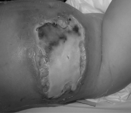 [Figure caption and citation for the preceding image starts]: 一名 2 岁女孩水痘感染后右腹部继发出现坏死性筋膜炎。来源于:de Benedictis FM, Osimani P. Necrotising fasciitis complicating varicella. BMJ Case Rep. 2009;2009:bcr2008141994 [Citation ends].
 [Figure caption and citation for the preceding image starts]: 坏死性筋膜炎的晚期体征包括广泛性蜂窝织炎、皮肤硬化、坏死以及出血性大疱形成。来源于:Hasham S, Matteucci P, Stanley PRW, et al. Necrotising fasciitis. BMJ. 2005 Apr 9;330(7495):830-3 [Citation ends].
强烈建议咨询传染病专家,以协助制定经验性抗生素治疗方案以及随后适当的降级治疗方案。
