多数 Paget 病患者没有症状。有症状的患者最初通常被误诊为其他肌肉骨骼疾病。通常是在患者进行针对其他诊断(例如检查肾疾病)的诊断性检查时,在影像学检查中偶然发现本病,并且仅用实验室或 X 线检查结果就可以澄清诊断。
临床发现
患者通常没有症状,但可能表现为长骨严重疼痛,罕见情况下可表现为面部区域的疼痛。疼痛可能继发于受累骨变弱导致的病理性骨折,最常见于股骨/髋骨或胫骨。基础病变可能表现为长骨的严重畸形(例如弯曲),这种症状也可能发生在扁骨,尤其是颅骨。这可能表现为明显的额部隆起或下颌前突。面骨受累时,可能会出现牙齿松动或咀嚼障碍。局部体温可能升高,原因是代谢活动增加。
患者可能表现有听力受损,为颞骨岩部受累所致。
由于存在椎管变窄或者盗血机制,这些患者常有椎管狭窄的并发症。 这可能表现为感觉异常和肌无力。[27]Parvizi J, Restrepo C, Sim FH. Paget's disease of the hip: surgical management. Future Rheumatology. 2006;1:373-7.[28]Yost JH, Spencer-Green G, Krant JD. Vascular steal mimicking compression myelopathy in Paget's disease of bone: rapid reversal with calcitonin and systemic steroids. J Rheumatol. 1993 Jun;20(6):1064-5.http://www.ncbi.nlm.nih.gov/pubmed/8350315?tool=bestpractice.com [Figure caption and citation for the preceding image starts]: 佩吉特病弯曲的胫骨(军刀状胫骨)宾夕法尼亚州费城罗思曼学院 Camilo Restrepo 提供 [Citation ends].
初始影像学检查
X 线平片
X 光检查对佩吉特病的评估极为重要。 所有患者在初始评估时均进行 X 线检查。[29]Singer FR, Bone HG 3rd, Hosking DJ, et al. Paget's disease of bone: an Endocrine Society clinical practice guideline. J Clin Endocrinol Metab. 2014 Dec;99(12):4408-22.http://press.endocrine.org/doi/full/10.1210/jc.2014-2910http://www.ncbi.nlm.nih.gov/pubmed/25406796?tool=bestpractice.com Paget骨病在 X 线片上有典型外观。
在疾病的后期,X 线检查主要表现为骨硬化,而不是溶骨性改变。所有三期病变可能会同时出现于同一患者的同一处骨骼中。作为代偿,新骨形成,X 光平片上可见特征性增厚的粗糙的骨小梁,对应组织学上表现为结构杂乱的板层骨,呈“马赛克”样。[27]Parvizi J, Restrepo C, Sim FH. Paget's disease of the hip: surgical management. Future Rheumatology. 2006;1:373-7.
骨扫描(例如闪烁图)
除了监测对旨在减少佩吉特病变部位放射性核素摄取的治疗反应外,不推荐进行 Tc-双膦酸盐连续扫描。[29]Singer FR, Bone HG 3rd, Hosking DJ, et al. Paget's disease of bone: an Endocrine Society clinical practice guideline. J Clin Endocrinol Metab. 2014 Dec;99(12):4408-22.http://press.endocrine.org/doi/full/10.1210/jc.2014-2910http://www.ncbi.nlm.nih.gov/pubmed/25406796?tool=bestpractice.com[31]Avramidis A, Polyzos SA, Moralidis E, et al. Scintigraphic, biochemical, and clinical response to zoledronic acid treatment in patients with Paget's disease of bone. J Bone Miner Metab. 2008;26(6):635-41.http://www.ncbi.nlm.nih.gov/pubmed/18979164?tool=bestpractice.com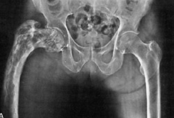 [Figure caption and citation for the preceding image starts]: 佩吉特病股骨近端X线片宾夕法尼亚州费城罗思曼学院 Camilo Restrepo 提供 [Citation ends].
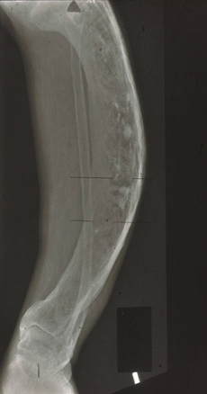 [Figure caption and citation for the preceding image starts]: 佩吉特病胫骨X线片宾夕法尼亚州费城罗思曼学院 Camilo Restrepo 提供 [Citation ends].
实验室检测
当怀疑患者患有佩吉特病时,则对其进行以下所有检查。
血清总碱性磷酸酶是骨形成的主要标志物,也是成骨细胞活动的指标之一。 血清碱性磷酸酶通常会升高,但佩吉特病患者的碱性磷酸酶也可能正常 (15%)。[32]Delmas PD, Meunier PJ. The management of Paget's disease of bone. N Engl J Med. 1997 Feb 20;336(8):558-66.http://www.ncbi.nlm.nih.gov/pubmed/9023094?tool=bestpractice.com 然而一项研究发现,高达 86% 的患者的碱性磷酸酶水平正常。[33]Eekhoff ME, van der Klift M, Kroon HM, et al. Paget's disease of bone in The Netherlands: a population-based radiological and biochemical survey--The Rotterdam Study. J Bone Miner Res. 2004 Apr;19(4):566-70.http://www.ncbi.nlm.nih.gov/pubmed/15005843?tool=bestpractice.com 碱性磷酸酶水平升高时,可以据此来监测治疗反应。[29]Singer FR, Bone HG 3rd, Hosking DJ, et al. Paget's disease of bone: an Endocrine Society clinical practice guideline. J Clin Endocrinol Metab. 2014 Dec;99(12):4408-22.http://press.endocrine.org/doi/full/10.1210/jc.2014-2910http://www.ncbi.nlm.nih.gov/pubmed/25406796?tool=bestpractice.com
骨特异性碱性磷酸酶是一种更敏感的诊断用血液检查,也可用作判断治疗反应的一个指标,但其普及性可能有限。[34]Alvarez L, Guanabens N, Peris P, et al. Discriminative value of biochemical markers of bone turnover in assessing the activity of Paget's disease. J Bone Miner Res. 1995 Mar;10(3):458-65.http://www.ncbi.nlm.nih.gov/pubmed/7785468?tool=bestpractice.com
血清前胶原 1 N-端肽(procollagen 1 N-terminal peptide, P1NP;I 型胶原的氨基端前肽)是骨形成的特异性衡量指标,其在骨闪烁显像上显示的活性与骨形成密切相关,[29]Singer FR, Bone HG 3rd, Hosking DJ, et al. Paget's disease of bone: an Endocrine Society clinical practice guideline. J Clin Endocrinol Metab. 2014 Dec;99(12):4408-22.http://press.endocrine.org/doi/full/10.1210/jc.2014-2910http://www.ncbi.nlm.nih.gov/pubmed/25406796?tool=bestpractice.com 但普及性可能有限且/或受到成本过高的限制。P1NP 对血清碱性磷酸酶可能正常的单骨性疾病或局限性疾病患者可能特别有用。P1NP 也可用作监测治疗反应的指标。
在未经治疗的佩吉特病患者中,I 型胶原蛋白的血清 C-端前肽(CTX,骨吸收活性的一个衡量指标)可能会升高。 血清 CTX 可用于监测治疗反应,因为经双膦酸盐治疗后,该指标会迅速下降。[29]Singer FR, Bone HG 3rd, Hosking DJ, et al. Paget's disease of bone: an Endocrine Society clinical practice guideline. J Clin Endocrinol Metab. 2014 Dec;99(12):4408-22.http://press.endocrine.org/doi/full/10.1210/jc.2014-2910http://www.ncbi.nlm.nih.gov/pubmed/25406796?tool=bestpractice.com
通过测量血清 25-羟基维生素 D 水平,可排除碱性磷酸酶升高的另一病因——骨软化症。 佩吉特病的血清 25-羟基维生素 D 水平正常。
应检查其他肝功能项目,以评估可引起碱性磷酸酶增高的肝病。
推荐进行血钙测量。应像对其他患者一样对 Paget 病患者进行高钙血症检查,但只有在极少数情况下,Paget 病可能会引发高钙血症,尤其是长期卧床休息后。
进一步影像学检查
计算机体层成像 (CT) 扫描和磁共振成像 (MRI) 主要用于评估非典型表现。[27]Parvizi J, Restrepo C, Sim FH. Paget's disease of the hip: surgical management. Future Rheumatology. 2006;1:373-7.[30]Whitehouse RW. Paget's disease of bone. Semin Musculoskelet Radiol. 2002 Dec;6(4):313-22.http://www.ncbi.nlm.nih.gov/pubmed/12541188?tool=bestpractice.com 当患者出现神经系统受累时,影像学检查可确定神经系统症状是否与 paget 样骨的代谢性和生理性骨改变有关。[35]Rubin DJ, Levin RM. Neurologic complications of Paget disease of bone. Endocr Pract. 2009 Mar;15(2):158-66.http://www.ncbi.nlm.nih.gov/pubmed/19289329?tool=bestpractice.com 影像学检查还用于检查可能的恶化。 paget样骨在横断面上(例如 CT 和 MRI 扫描)看起来宽大且排列无序。
骨活检
骨活检最敏感且最特异,但很少需要。 承重长骨,例如股骨,应避免使用诊断性活检,因为可能导致损伤骨出现骨折。[36]Eisman JA, Martin TJ. Osteolytic Paget's disease: recognition and risks of biopsy. J Bone Joint Surg Am. 1986 Jan;68(1):112-7.http://www.ncbi.nlm.nih.gov/pubmed/3941112?tool=bestpractice.com 如果在所有初始检查之后诊断仍有疑问,则要求进行活检。
