脊柱包括 2 类基本的软骨关节:椎间盘(主要含有复合胶冻状物质)以及面关节(滑膜关节)。[8]Rao RD, Currier BL, Albert TJ, et al. Degenerative cervical spondylosis: clinical syndromes, pathogenesis and management. J Bone Joint Surg Am. 2007;89:1360-1378.http://www.ncbi.nlm.nih.gov/pubmed/17575617?tool=bestpractice.com
由于椎间盘主要细胞(维持胶冻样)的丢失以及终板的硬化(防止营养物质的弥散),椎间盘胶冻样物质随着成熟很难维持。椎间盘关节变得脱水而狭窄。椎间盘的环通常没有神经分布,但在一定程度的狭窄时,可出现神经分布,并在边缘发展为骨赘,与任一活动关节类似。由于面关节功能主要是防止旋转、弯曲/伸展,面关节退化的进展是由于椎间盘关节的狭窄所产生的更多的轴向负重,从而施压于关节突关节。
在大多数人群中,颈椎关节的退行性变或椎关节强硬是无症状的,部分颈椎活动度减小的患者除外。[1]Matsumoto M, Fujimura Y, Suzuki N, et al. MRI of cervical intervertebral discs in asymptomatic subjects. J Bone Joint Surg Br. 1998;80:19-24.http://www.bjj.boneandjoint.org.uk/content/80-B/1/19.full.pdf+htmlhttp://www.ncbi.nlm.nih.gov/pubmed/9460946?tool=bestpractice.com[4]Siivola SM, Levoska S, Tervonen O, et al. MRI changes of cervical spine in asymptomatic and symptomatic young adults. Eur Spine J. 2002;11:358-363.http://www.ncbi.nlm.nih.gov/pubmed/12193998?tool=bestpractice.com
然而,一部分患者会出现伴随轻度颈椎退行性病变的轴颈痛(例如仅表现为局限性关节狭窄)。[16]Haldeman S, Carroll L, Cassidy JD. Findings from the bone and joint decade 2000 to 2010 task force on neck pain and its associated disorders. J Occup Environ Med. 2010;52:424-427.http://www.ncbi.nlm.nih.gov/pubmed/20357682?tool=bestpractice.com因此,颈椎平片或者 MRI 所示颈椎病并不一定与轴性颈椎痛的症状有关。[1]Matsumoto M, Fujimura Y, Suzuki N, et al. MRI of cervical intervertebral discs in asymptomatic subjects. J Bone Joint Surg Br. 1998;80:19-24.http://www.bjj.boneandjoint.org.uk/content/80-B/1/19.full.pdf+htmlhttp://www.ncbi.nlm.nih.gov/pubmed/9460946?tool=bestpractice.com[4]Siivola SM, Levoska S, Tervonen O, et al. MRI changes of cervical spine in asymptomatic and symptomatic young adults. Eur Spine J. 2002;11:358-363.http://www.ncbi.nlm.nih.gov/pubmed/12193998?tool=bestpractice.com[17]Gross AR, Goldsmith C, Hoving JL, et al.; Cervical Overview Group. Conservative management of mechanical neck disorders: a systematic review. J Rheumatol. 2007;34:1083-1102.http://www.ncbi.nlm.nih.gov/pubmed/17295434?tool=bestpractice.com 颈椎病疼痛类型的感受主要来自投射至颈部脊椎旁肌肉的关节受体信号(包括支配退化纤维环的神经分布异常),导致脊旁肌肉痉挛以及肩胛间区和颈外侧疼痛。[2]Guzman J, Haldeman S, Carroll LJ, et al. Clinical practice implications of the Bone and Joint Decade 2000-2010 Task Force on Neck Pain and Its Associated Disorders: from concepts and findings to recommendations. J Manipulative Physiol Ther. 2009;32(2 Suppl):S227-S243.http://www.ncbi.nlm.nih.gov/pubmed/19251069?tool=bestpractice.com[6]Binder AI. Neck pain. BMJ Clin Evid. 2008. http://clinicalevidence.bmj.com/ (last accessed 9 August 2016).http://www.ncbi.nlm.nih.gov/pmc/articles/PMC2907992/http://www.ncbi.nlm.nih.gov/pubmed/19445809?tool=bestpractice.com[7]Binder AI. Cervical spondylosis and neck pain. BMJ. 2007;334:527-531.http://www.ncbi.nlm.nih.gov/pubmed/17347239?tool=bestpractice.com 退行性变伴发的自发性轴颈痛,比特殊原因引起的轴颈痛预后更差。[18]Hush JM. Prognosis of acute idiopathic neck pain is poor: a systematic review and meta-analysis. Arch Phys Med Rehabil. 2011;92:824-829.http://www.ncbi.nlm.nih.gov/pubmed/21458776?tool=bestpractice.com
神经根型颈椎病 (CSR) 是以下两种情况的结果:在颈神经出脊髓时椎管受到退行椎间盘压迫(即:椎间盘突出,此处椎间盘的纤维环薄弱导致髓核内容物移位至临近神经根);颈椎发生中至重度的退行性变导致椎间孔神经根出口狭窄,引起神经根压迫。[19]Van Zundert J, Huntoon M, Patijn J, et al. 4. Cervical radicular pain. Pain Pract. 2010;10:1-17.http://www.ncbi.nlm.nih.gov/pubmed/19807874?tool=bestpractice.com[20]Bono CM, Ghiselli G, Gilbert TJ, et al; North American Spine Society. An evidence-based clinical guideline for the diagnosis and treatment of cervical radiculopathy from degenerative disorders. Spine J. 2011;11:64-72.http://www.ncbi.nlm.nih.gov/pubmed/21168100?tool=bestpractice.com
脊髓型颈椎病 (CSM) 通常包括严重椎间盘以及面关节的退行性变伴有脊椎对位方式的改变,例如脊柱后凸或前移、伴有骨赘形成等。这些可能导致严重的椎管狭窄和继发性脊髓变形。[12]Rao RD, Gourab K, David KS. Operative treatment of cervical spondylotic myelopathy. J Bone Surg Am. 2006;88:1619-1640.http://www.ncbi.nlm.nih.gov/pubmed/16818991?tool=bestpractice.com [Figure caption and citation for the preceding image starts]: 颈椎 MRI(矢状位 T2)显示中度退行性关节病,但无明显脊髓压迫Dennis A. Turner, MA, MD [Citation ends].
 [Figure caption and citation for the preceding image starts]: 颈椎 MRI(矢状位 T2)显示轻度退行性关节病和椎间盘突出Dennis A. Turner, MA, MD [Citation ends].
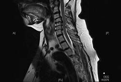 [Figure caption and citation for the preceding image starts]: 颈椎 MRI 显示(矢状位 T2 像):严重多节段椎间盘退行性变,但无明显脊髓受压(即:无畸形,也无内在 T2 改变)Dennis A. Turner, MA, MD [Citation ends].
 [Figure caption and citation for the preceding image starts]: 有症状的脊髓型颈椎病患者的颈椎矢状位 T2 加权像显示单一节段的脊髓压迫Dennis A. Turner, MA, MD [Citation ends].
 [Figure caption and citation for the preceding image starts]: 矢状位 T2 MRI 显示前期 C3/4 水平脊髓受压,伴有残留 T2 改变;在 C2/3 和 C6/7 出现新的脊髓压迫,T2 改变Dennis A. Turner, MA, MD [Citation ends].
 [Figure caption and citation for the preceding image starts]: 颈椎病分类表,包括无症状(放射影像学上)颈椎病可能出现的各种症状Dennis A. Turner, MA, MD [Citation ends].