案例#1
一名 55 岁女性因右胁肿块前来就诊。 患者陈述她最近被诊断出糖尿病,但她已能通过饮食改变控制糖尿病。 她在 3 个月内体重减轻 9 kg(20 磅),随后发现其右下胸腔外侧有一个肿块。 无痛,但主诉穿慢跑胸罩时会觉得不适。 体格检查发现肿块柔软、浅表性、且可移动,直径为 5 cm。
案例#2
一名 35 岁的男性因右大腿结节和之前瘢痕处复发性左胸壁结节前来就诊。 患者陈述他 2 年前发现其右大腿外侧有一肿块,左胸壁病变已于 3 年前在诊所去除。 最近数月,结节略有增大。 他还陈述,他碰触这些结节时会有不适感。 体格检查发现肿块大小为 1 cm x 2 cm,柔软、可移动且位于皮下。
其他表现
除了躯干或四肢近端皮下组织,脂肪瘤还可出现在其他部位。胃肠道脂肪瘤为黏膜下病变,最常见于食管、胃部和小肠。这种类型可表现为肠梗阻或出血。罕见情况下,脂肪瘤还可发生于肾上腺、腮腺、咽旁间隙、乳房、纵隔膜、胸膜、大气道、心脏、上腔静脉、脑部及脊柱内等部位。
家族多发性脂肪过多症患者,还会因遗传原因而发生。[8]Leffell DJ, Braverman IM. Familial multiple lipomatosis: report of a case and a review of the literature. J Am Acad Dermatol. 1986;15:275-279.http://www.ncbi.nlm.nih.gov/pubmed/3745530?tool=bestpractice.com[9]Toy BR. Familial multiple lipomatosis. Dermatol Online J. 2003;9:9.http://www.ncbi.nlm.nih.gov/pubmed/14594582?tool=bestpractice.com 有这种常染色体遗传性疾病的患者往往是男性,并且此类患者存在四肢和躯干多发性、广泛性、对称性脂肪瘤。[2]Koh HK, Bhawan J. Tumors of the skin. In: Moschella SL, Hurley HJ, eds. Dermatology. 3rd ed. Philadelphia: WB Saunders. 1992:1721-1808.[10]Enzi G. Multiple symmetric lipomatosis: an updated clinical report. Medicine (Baltimore). 1984;63:56-64.http://www.ncbi.nlm.nih.gov/pubmed/6318013?tool=bestpractice.com 其他与脂肪瘤有关的遗传性综合征包括 Madelung 病,该病与重度饮酒男性有关,特征为头、颈、肩及近端上肢良性对称性脂肪过多症;[2]Koh HK, Bhawan J. Tumors of the skin. In: Moschella SL, Hurley HJ, eds. Dermatology. 3rd ed. Philadelphia: WB Saunders. 1992:1721-1808.[11]Uhlin SR. Benign symmetric lipomatosis. Arch Dermatol. 1979;115:94-95.http://www.ncbi.nlm.nih.gov/pubmed/760666?tool=bestpractice.com 还包括德尔肯氏病(也被称为痛性肥胖病),该病发生于中年女性,特征为躯干、肩部、手臂及腿部疼痛性脂肪瘤。[12]Wortham NC, Tomlinson JP. Dercum's disease. Skinmed. 2005;4:157-162.http://www.ncbi.nlm.nih.gov/pubmed/15891252?tool=bestpractice.com
血管脂肪瘤表现为疼痛性皮下小结,通常发生于青年人群,且 50% 以上的病例为多发。[2]Koh HK, Bhawan J. Tumors of the skin. In: Moschella SL, Hurley HJ, eds. Dermatology. 3rd ed. Philadelphia: WB Saunders. 1992:1721-1808.[3]Austin RM, Mack GR, Townsend CM, et al. Infiltrating (intramuscular) lipomas and angiolipomas: a clinicopathologic study of six cases. Arch Surg. 1980;115:281-284.http://www.ncbi.nlm.nih.gov/pubmed/7356383?tool=bestpractice.com[13]Dalal KM, Antonescu CR, Singer S. Diagnosis and management of lipomatous tumors. J Surg Oncol. 2008;97:298-313.http://www.ncbi.nlm.nih.gov/pubmed/18286473?tool=bestpractice.com 其中的脂肪细胞散布着包含纤维蛋白血栓的集群毛细血管。
梭形细胞脂肪瘤,常见于 45-65 岁男性,易发于颈后和肩部区。[13]Dalal KM, Antonescu CR, Singer S. Diagnosis and management of lipomatous tumors. J Surg Oncol. 2008;97:298-313.http://www.ncbi.nlm.nih.gov/pubmed/18286473?tool=bestpractice.com 主要特征为形成胶原的梭形细胞代替成熟脂肪。[14]Fanburg-Smith JC, Devaney KO, Miettinen M, et al. Multiple spindle cell lipomas: a report of 7 familial and 11 nonfamilial cases. Am J Surg Pathol. 1998;22:40-48.http://www.ncbi.nlm.nih.gov/pubmed/9422314?tool=bestpractice.com[15]Brody HJ, Meltzer HD, Someren A. Spindle cell lipoma: an unusual dermatologic presentation. Arch Dermatol. 1978;114:1065-1066.http://www.ncbi.nlm.nih.gov/pubmed/686729?tool=bestpractice.com
肌内脂肪瘤通常边界不清且具有浸润性,一般见于中年人群,表现为大腿或躯干部缓慢生长的深部肿块。排除不典型脂肪瘤性肿瘤或高分化脂肪肉瘤非常重要,因为在这些解剖部位,这些肿瘤比肌内脂肪瘤更常见。[13]Dalal KM, Antonescu CR, Singer S. Diagnosis and management of lipomatous tumors. J Surg Oncol. 2008;97:298-313.http://www.ncbi.nlm.nih.gov/pubmed/18286473?tool=bestpractice.com[16]Kooby DA, Antonescu CR, Brennan MF, et al. Atypical lipomatous tumor/well-differentiated liposarcoma of the extremity and trunk wall: importance of histological subtype with treatment recommendations. Ann Surg Oncol. 2004;11:78-84.http://www.ncbi.nlm.nih.gov/pubmed/14699038?tool=bestpractice.com 在腹膜后病变病例中也应考虑这些诊断。
蛰伏脂瘤可发生于躯干、腹膜后及四肢,类似于冬眠动物的腺状褐色脂肪。[5]Ahn C, Harvey JC. Mediastinal hibernoma, a rare tumor. Ann Thorac Surg. 1990;50:828-830.http://www.ncbi.nlm.nih.gov/pubmed/2241353?tool=bestpractice.com[13]Dalal KM, Antonescu CR, Singer S. Diagnosis and management of lipomatous tumors. J Surg Oncol. 2008;97:298-313.http://www.ncbi.nlm.nih.gov/pubmed/18286473?tool=bestpractice.com 手术期间容易出血,如果不完整切除肿瘤,术后容易复发。 [Figure caption and citation for the preceding image starts]: 躯干皮下脂肪瘤来自 Kimberly Moore Dalal 博士和 Steven D. DeMartini 博士收集的资料;经许可后使用 [Citation ends].
 [Figure caption and citation for the preceding image starts]: 胃黏膜下脂肪瘤,CT 扫描。 遍布整个黏膜下窦的脂肪密度肿块。来自 Kimberly Moore Dalal 博士和 Steven D. DeMartini 博士收集的资料;经许可后使用 [Citation ends].
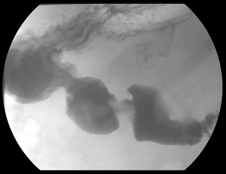 [Figure caption and citation for the preceding image starts]: 胃黏膜下脂肪瘤,上消化道造影检查。 末端腔和幽门通道中的充盈缺损提示窦肿块脱入幽门通道中来自 Kimberly Moore Dalal 博士和 Steven D. DeMartini 博士收集的资料;经许可后使用 [Citation ends].
 [Figure caption and citation for the preceding image starts]: 胃黏膜下脂肪瘤。 成熟脂肪组织结节位于胃黏膜下方。 苏木精 - 伊红染色法,放大倍率为 20 倍来自 Kimberly Moore Dalal 博士和 Steven D. DeMartini 博士收集的资料;经许可后使用 [Citation ends].
 [Figure caption and citation for the preceding image starts]: 血管脂肪瘤。 成熟的脂肪组织(含内皮细胞增生的微血管血栓灶)。 苏木精 - 伊红染色法,放大倍率为 200 倍来自 Kimberly Moore Dalal 博士和 Steven D. DeMartini 博士收集的资料;经许可后使用 [Citation ends].
 [Figure caption and citation for the preceding image starts]: 梭形细胞脂肪瘤。 成熟脂肪组织(中间有含梭形细胞区和典型的旧胶原蛋白束的密度纤维化条索)。 苏木精 - 伊红染色法,放大倍率为 200 倍来自 Kimberly Moore Dalal 博士和 Steven D. DeMartini 博士收集的资料;经许可后使用 [Citation ends].
 [Figure caption and citation for the preceding image starts]: 肌内脂肪瘤,右大腿。 轴向 T1 加权 MR 图像。 位于右大腿前面部位的脂肪瘤性肿块来自 Kimberly Moore Dalal 博士和 Steven D. DeMartini 博士收集的资料;经许可后使用 [Citation ends].
 [Figure caption and citation for the preceding image starts]: 肌内脂肪瘤,右大腿。 冠状面 T1 加权 MRI 图像。 位于右大腿前面部位的脂肪瘤性肿块来自 Kimberly Moore Dalal 博士和 Steven D. DeMartini 博士收集的资料;经许可后使用 [Citation ends].
 [Figure caption and citation for the preceding image starts]: 肩胛下肌肌内脂肪瘤,CT 扫描。 边界清楚的右腋窝软组织脂肪肿块来自 Kimberly Moore Dalal 博士和 Steven D. DeMartini 博士收集的资料;经许可后使用 [Citation ends].
 [Figure caption and citation for the preceding image starts]: 肌内脂肪瘤。 骨骼肌束间的成熟脂肪组织。 苏木精 - 伊红染色法,放大倍率为 200 倍来自 Kimberly Moore Dalal 博士和 Steven D. DeMartini 博士收集的资料;经许可后使用 [Citation ends].
