识别患者临床类别(艾滋病相关性、经典散发性、移植相关性或非洲地方性)和病变外观,将有助于正确诊断 KS 的首发病变。通过活检和组织病理学确诊。[17]Radu O, Pantanowitz L. Kaposi sarcoma. Arch Pathol Lab Med. 2013 Feb;137(2):289-94.http://www.archivesofpathology.org/doi/full/10.5858/arpa.2012-0101-RShttp://www.ncbi.nlm.nih.gov/pubmed/23368874?tool=bestpractice.com [Figure caption and citation for the preceding image starts]: 显示成血管梭形瘤细胞束(苏木精和伊红染色)的卡波西肉瘤组织病理学显微照片由 Liron Pantanowitz 博士提供;经许可后使用 [Citation ends].
病史
患者可能报告出现皮肤病损,经常伴有心理社会应激(焦虑)。皮肤病变通常无痛且不痒,因此也可能不出现症状。小腿上的肿瘤可能影响步态。腿、生殖器和面部的淋巴水肿可能有疼痛感。口腔损害可能出现出血和溃疡,并且可能影响咀嚼、言语和吞咽。胃肠道卡波西肉瘤可能引起体重减轻、腹痛、恶心和呕吐、出血或腹泻。肺部 KS 可能表现为呼吸困难、发热、咳嗽、咯血或胸痛。在表现为基础疾病(例如 HIV 感染或移植)或者因基础疾病接受随访的患者中,KS 也可能是一种偶然发现。
人疱疹病毒 8 (HHV-8) 感染是 KS 发展的必然因素。 患有其他 HHV-8 相关疾病(例如,多中心 Castleman 病的患者和原发性渗出性淋巴瘤 (PEL))的患者也有发生伴随性 KS 的风险。[13]Sullivan RJ, Pantanowitz L, Casper C, et al. HIV/AIDS: epidemiology, pathophysiology, and treatment of Kaposi sarcoma-associated herpesvirus disease: Kaposi sarcoma, primary effusion lymphoma, and multicentric Castleman disease. Clin Infect Dis. 2008 Nov 1;47(9):1209-15.https://www.ncbi.nlm.nih.gov/pmc/articles/PMC2700291/http://www.ncbi.nlm.nih.gov/pubmed/18808357?tool=bestpractice.com [Figure caption and citation for the preceding image starts]: 下肢上的多个粉紫色卡波西肉瘤结节由 Bruce J. Dezube 博士提供;经许可后使用 [Citation ends].
查体
KS 患者的初步评估包括全身体格检查,特别注意经常出现病变损害的区域,例如下肢、面部、口腔黏膜和生殖器。[14]Dezube BJ, Pantanowitz L, Aboulafia DM. Management of AIDS-related Kaposi sarcoma: advances in target discovery and treatment. AIDS Read. 2004 May;14(5):236-8.http://www.ncbi.nlm.nih.gov/pubmed/15199854?tool=bestpractice.com 应将所有皮肤黏膜病变记录在案。
KS 皮肤病损通常具有以下特点:
多灶性
非对称性分布
大小不一
颜色多样(粉红色、红色、紫色、棕色或蓝色)
结节性[Figure caption and citation for the preceding image starts]: 下肢上的多个粉紫色卡波西肉瘤结节由 Bruce J. Dezube 博士提供;经许可后使用 [Citation ends].
丘疹型
斑块样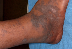 [Figure caption and citation for the preceding image starts]: 足部卡波西肉瘤皮肤紫褐色斑块由 Bruce J. Dezube 博士提供;经许可后使用 [Citation ends].
大疱状
硬结(木质)
疣状
蕈伞型
溃疡性
感染性。
淋巴水肿可能比较广泛,因此可能掩盖底层的 KS。 应确定是否有内脏受累,有或无皮肤黏膜损伤,应特别注意淋巴结、GI 道和呼吸系统。
检查
在出现 KS 的人群中应进行艾滋病病毒检查。 若为 HIV 阳性,则应获得 CD4 T-细胞计数和艾滋病病毒的病毒负荷量。 利用小钻取2~4 mm 临床疑似皮肤病损活检进行组织病理学确诊。 皮肤钻取活检易于执行,并可随时帮助鉴别诊断。 病理组织学可见具特征性的非典型梭形细胞。[17]Radu O, Pantanowitz L. Kaposi sarcoma. Arch Pathol Lab Med. 2013 Feb;137(2):289-94.http://www.archivesofpathology.org/doi/full/10.5858/arpa.2012-0101-RShttp://www.ncbi.nlm.nih.gov/pubmed/23368874?tool=bestpractice.comKS 患者的病变细胞表达 HHV-8 潜核抗原-1 (LNA-1)(可使用免疫组化检测)、血管内皮细胞免疫标记物(例如 CD31 和 CD34)和淋巴细胞免疫标记物(例如 D2-40)。[18]Cheuk W, Wong KO, Wong CS, et al. Immunostaining for human herpesvirus 8 latent nuclear antigen-1 helps distinguish Kaposi sarcoma from its mimickers. Am J Clin Pathol. 2004 Mar;121(3):335-42.http://www.ncbi.nlm.nih.gov/pubmed/15023037?tool=bestpractice.com[19]Patel RM, Goldblum JR, Hsi ED. Immunohistochemical detection of human herpesvirus-8 latent nuclear antigen-1 is useful in the diagnosis of Kaposi sarcoma. Mod Pathol. 2004 Apr;17(4):456-60.http://www.nature.com/modpathol/journal/v17/n4/full/3800061a.htmlhttp://www.ncbi.nlm.nih.gov/pubmed/14990970?tool=bestpractice.com 因为大块淋巴结肿大包括若干实体,故在这种情况下可能需要诊断活检。
利用胸部 X 射线 (CXR) 或 CT 扫描的肺部成像有益于筛查呼吸道症状患者是否患有肺 KS。 必须排除伴发性肺感染。 使用支气管镜检查确定肺KS诊断适用于存在胸片异常的患者以及已证实的肺受累致使治疗方案改变的患者。[14]Dezube BJ, Pantanowitz L, Aboulafia DM. Management of AIDS-related Kaposi sarcoma: advances in target discovery and treatment. AIDS Read. 2004 May;14(5):236-8.http://www.ncbi.nlm.nih.gov/pubmed/15199854?tool=bestpractice.com 由于存在出血风险,呼吸道病变活检存在相对禁忌。[20]Pitchenik AE, Fischl MA, Saldana MJ. Kaposi's sarcoma of the tracheobronchial tree. Clinical, bronchoscopic, and pathologic features. Chest. 1985 Jan;87(1):122-4.http://www.ncbi.nlm.nih.gov/pubmed/3965256?tool=bestpractice.com[21]Zibrak JD, Silvestri RC, Costello P, et al. Bronchoscopic and radiologic features of Kaposi's sarcoma involving the respiratory system. Chest. 1986 Oct;90(4):476-9.http://www.ncbi.nlm.nih.gov/pubmed/3489584?tool=bestpractice.com[22]Kodra A, Walczyszyn M, Grossman C, et al. Case report: pulmonary Kaposi sarcoma in a non-HIV patient. F1000Res. 2015 Oct 7;4:1013.https://f1000research.com/articles/4-1013/v1http://www.ncbi.nlm.nih.gov/pubmed/26664711?tool=bestpractice.com
人疱疹病毒 8 (HHV-8) 血清学可能指示患者处于 KS 病负担的风险和/或与其相关。 这些检查的临床实用性需进一步验证。[23]Casper C, Krantz E, Taylor H, et al. Assessment of a combined testing strategy for detection of antibodies to human herpesvirus 8 (HHV-8) in persons with Kaposi's sarcoma, persons with asymptomatic HHV-8 infection, and persons at low risk for HHV-8 infection. J Clin Microbiol. 2002 Oct;40(10):3822-5.http://jcm.asm.org/content/40/10/3822.fullhttp://www.ncbi.nlm.nih.gov/pubmed/12354890?tool=bestpractice.com
大便潜血对于筛查 GI 道疾病有帮助。[14]Dezube BJ, Pantanowitz L, Aboulafia DM. Management of AIDS-related Kaposi sarcoma: advances in target discovery and treatment. AIDS Read. 2004 May;14(5):236-8.http://www.ncbi.nlm.nih.gov/pubmed/15199854?tool=bestpractice.com 对于有症状的个体患者,GI 内窥镜检查可能有帮助。CT 扫描、PET 扫描和 MRI 检查可能有助于评估深部结节性和脏器 KS。CT 扫描和 MRI 能够更好地检测骨病变,因为普通 X 线片或骨扫描往往不能检测到。[24]Caponetti G, Dezube BJ, Restrepo C, et al. Kaposi sarcoma of the musculoskeletal system: a review of 66 patients. Cancer. 2007 Mar 15;109(6):1040-52.http://onlinelibrary.wiley.com/doi/10.1002/cncr.22500/fullhttp://www.ncbi.nlm.nih.gov/pubmed/17265518?tool=bestpractice.com 在需全身化疗的患者中需评估肝、肾和骨髓功能。[Figure caption and citation for the preceding image starts]: 显示成血管梭形瘤细胞束(苏木精和伊红染色)的卡波西肉瘤组织病理学显微照片由 Liron Pantanowitz 博士提供;经许可后使用 [Citation ends]. [Figure caption and citation for the preceding image starts]: 显示网状的胸部 CT 扫描(因卡波西肉瘤导致肺受累)由 Bruce J. Dezube 博士提供;经许可后使用 [Citation ends].
