须根据病史和临床发现进行诊断。影像学检查并非诊断所必需,但其在评估疾病发展和治疗规划方面具有重要价值。
病史和检查
患者通常表现为听力下降或耳鸣。后天性胆脂瘤中常见的是,具有复发性或慢性化脓性耳分泌物的病史或表现,可能对抗生素治疗无反应。分泌物恶臭,但可能不多。少见的是,患者出现耳痛、眩晕或面神经 (VII) 功能障碍(味觉改变或面神经无力)等症状。这些往往预示着病情已经进展。
应询问患者之前是否存在任何耳部疾病或咽鼓管功能障碍的病因。[4]Semaan MT, Megerian CA. The pathophysiology of cholesteatoma. Otolaryngol Clin North Am. 2006 Dec;39(6):1143-59.http://www.ncbi.nlm.nih.gov/pubmed/17097438?tool=bestpractice.com[23]Isaacson G. Diagnosis of pediatric cholesteatoma. Pediatrics. 2007 Sep;120(3):603-8.http://www.ncbi.nlm.nih.gov/pubmed/17766534?tool=bestpractice.com 伴发颚裂、颅面畸形、特纳综合征或唐氏综合征,可增加后天性胆脂瘤的风险。[14]Kemppainen HO, Puhakka HJ, Laippala PJ, et al. Epidemiology and aetiology of middle ear cholesteatoma. Acta Otolaryngol. 1999;119(5):568-72.http://www.ncbi.nlm.nih.gov/pubmed/10478597?tool=bestpractice.com[18]Dhooge IJ, De Vel E, Verhoye C, et al. Otologic disease in Turner syndrome. Otol Neurotol. 2005 Mar;26(2):145-50.http://www.ncbi.nlm.nih.gov/pubmed/15793396?tool=bestpractice.com[19]Bacciu A, Pasanisi E, Vincenti V, et al. Surgical treatment of middle ear cholesteatoma in children with Down syndrome. Otol Neurotol. 2005 Sep;26(5):1007-10.http://www.ncbi.nlm.nih.gov/pubmed/16151350?tool=bestpractice.com 少见的是,家庭成员也可能患有中耳疾病或胆脂瘤。[23]Isaacson G. Diagnosis of pediatric cholesteatoma. Pediatrics. 2007 Sep;120(3):603-8.http://www.ncbi.nlm.nih.gov/pubmed/17766534?tool=bestpractice.com[31]Homoe P, Rosborg J. Family cluster of cholesteatoma. J Laryngol Otol. 2007 Jan;121(1):65-7.http://www.ncbi.nlm.nih.gov/pubmed/17059626?tool=bestpractice.com
检查时可能会发现耳分泌物的证据。耳镜检查通常发现上鼓室(中耳上部)、松弛部或紧张部(通常为后上方)有痂皮或角质,伴或不伴鼓膜穿孔。[32]Bhutta MF, Williamson IG, Sudhoff HH. Cholesteatoma. BMJ. 2011 Mar 3;342:d1088.http://www.ncbi.nlm.nih.gov/pubmed/21372073?tool=bestpractice.com [Figure caption and citation for the preceding image starts]: 上鼓室(中耳上部分)胆脂瘤来自 Susan Douglas 医生的个人教学集锦 [Citation ends].
 [Figure caption and citation for the preceding image starts]: 上鼓室内陷袋(中耳上部分)来自 Susan Douglas 医生的个人教学集锦 [Citation ends]. 对于先天性胆脂瘤,在完整的鼓膜后面可观察到白色肿块。
若有明显耳分泌物,则患者可能需用耳显微镜和微吸引器检查耳部。0° 或 30° 内窥镜也有助于耳部检查。有助于区分内陷袋和胆脂瘤。[33]Dement'eva NF, Shilenkov AA, Kozlov VS. Otoendoscopy for the diagnosis of chronic middle ear diseases. Vestn Otorinolaringol. 2010;(1):71-5.http://www.ncbi.nlm.nih.gov/pubmed/20436431?tool=bestpractice.com对于检查困难的患者(例如,小儿、学习困难者),可能需在全身麻醉下进行耳部检查。使其能够彻底吸除耳部分泌物、确定分泌物的原因(是否存在穿孔或内陷囊袋)以及有无胆脂瘤的存在。
有时进行瘘管试验,使用鼓膜测量技术施加正压,并记录眼球运动。施加正压后,阳性结果是眼球震颤。但是,试验可能给出假阴性结果。
听力学
应测听所有患者以确定其听力状态。听力测验结果可能正常,但更常见的是传导性听觉受损。耳蜗损伤或之前存在听力受损(例如,先天性或老年性耳聋)的患者中,可能出现兼有传导性和感音神经性成分的混合型听力受损。
影像学检查方法
建议将颞骨岩部高分辨率 CT 扫描作为中耳胆脂瘤患者初诊检查的一部分。[34]Ayache D, Darrouzet V, Dubrulle F, et al. Imaging of non-operated cholesteatoma: clinical practice guidelines. Eur Ann Otorhinolaryngol Head Neck Dis. 2012 Jun;129(3):148-52.http://www.ncbi.nlm.nih.gov/pubmed/22321912?tool=bestpractice.com可以明确具有非典型表现的患者病情,并可用于评估乳突的病理状态以及有无耳蜗、半规管或颅内侵入等并发症。[25]Hamilton J. Chronic otitis media in childhood. In: Gleeson M, ed. Scott-Brown's otorhinolaryngology: head and neck surgery. 7th ed. London: Edward Arnold Ltd; 2008:928-965.胆脂瘤患者CT 显示中耳或乳突混浊,也可能显示鼓室盾板、听骨链、迷路、面神经管、天盖或乙状窦骨板侵蚀。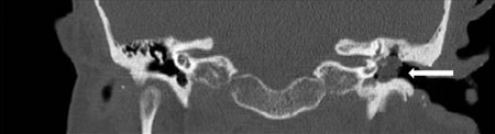 [Figure caption and citation for the preceding image starts]: 胆脂瘤,冠状 CT 扫描来自 Susan Douglas 医生的个人教学集锦 [Citation ends].
 [Figure caption and citation for the preceding image starts]: 胆脂瘤,轴位 CT 扫描来自 Susan Douglas 医生的个人教学集锦 [Citation ends].
MRI 对胆脂瘤的诊断有限。因其无法检查颞骨病变细节。但是,若怀疑有颅内并发症(例如,颞叶脓肿或脑膜炎)时,则有帮助。MRI可显示中耳和乳突混浊,或显示脑膜脑炎感染、颅内侵犯或乙状窦血栓形成等颅内并发症的迹象。[34]Ayache D, Darrouzet V, Dubrulle F, et al. Imaging of non-operated cholesteatoma: clinical practice guidelines. Eur Ann Otorhinolaryngol Head Neck Dis. 2012 Jun;129(3):148-52.http://www.ncbi.nlm.nih.gov/pubmed/22321912?tool=bestpractice.com已表明基于弥散加权磁共振成像 (MRI) 的非平面回波在已做过手术的患者中可用于排除疾病复发的可能。[35]De Foer B, Vercruysse JP, Bernaerts A, et al. Middle ear cholesteatoma: non-echo-planar diffusion-weighted MR imaging versus delayed gadolinium-enhanced T1-weighted MR imaging - value in detection. Radiology. 2010 Jun;255(3):866-72.http://www.ncbi.nlm.nih.gov/pubmed/20501723?tool=bestpractice.com[36]De Foer B, Vercruysse JP, Bernaerts A, et al. Detection of postoperative residual cholesteatoma with non-echo-planar diffusion-weighted magnetic resonance imaging. Otol Neurotol. 2008 Jun;29(4):513-7.http://www.ncbi.nlm.nih.gov/pubmed/18520587?tool=bestpractice.com[37]Li PM, Linos E, Gurgel RK, et al. Evaluating the utility of non-echo-planar diffusion-weighted imaging in the preoperative evaluation of cholesteatoma: a meta-analysis. Laryngoscope. 2013 May;123(5):1247-50.http://www.ncbi.nlm.nih.gov/pubmed/23023958?tool=bestpractice.com
微生物学
耳分泌物培养主要针对那些对抗菌治疗无反应的患者。耳拭子显示有细菌感染,通常为绿脓假单胞菌,也可能显示金黄色葡萄球菌和厌氧菌等其他病原体。[38]Brook I. Role of anaerobic bacteria in chronic otitis media and cholesteatoma. Int J Pediatr Otorhinolaryngol. 1995 Mar;31(2-3):153-7.http://www.ncbi.nlm.nih.gov/pubmed/7782173?tool=bestpractice.com
