世界卫生组织将口腔白斑病定义为口腔黏膜上以白色为主的损伤,该损伤并不能诊断为其他任何已明确的病变。[1]Kramer IR, Lucas RB, Pindborg JJ, et al. Definition of leukoplakia and related lesions: an aid to studies on oral precancer. Oral Surg Oral Med Oral Pathol. 1978;46:518-539.http://www.ncbi.nlm.nih.gov/pubmed/280847?tool=bestpractice.com[2]Axell T, Holmstrup P, Kramer IRH, et al. International seminar on oral leukoplakia and associated lesions related to tobacco habits. Community Dent Oral Epidemiol. 1984;12:145-154.[3]Axell T, Pindborg JJ, Smith CJ, et al. Oral white lesions with special reference to precancerous and tobacco-related lesions: conclusions of an international symposium held in Uppsala, Sweden, May 18-21 1994. International Collaborative Group on Oral White Lesions. J Oral Pathol Med. 1996;25:49-54.http://www.ncbi.nlm.nih.gov/pubmed/8667255?tool=bestpractice.com因此,其诊断需要排除其他所有可能。多数病损通过全面的病史采集和临床检查即可做出初步诊断。如果临床诊断不明确,可进行组织活检确诊。[51]Sankaranarayanan R, Fernandez Garrote L, Lence Anta J, et al. Visual inspection in oral cancer screening in Cuba: a case-control study. Oral Oncol. 2002;38:131-136.http://www.ncbi.nlm.nih.gov/pubmed/11854059?tool=bestpractice.com[52]Shugars DC, Patton LL. Detecting, diagnosing, and preventing oral cancer. Nurse Pract 1997;22:105, 109-110, 113-115.http://www.ncbi.nlm.nih.gov/pubmed/9211456?tool=bestpractice.com
尽管多数口腔白斑为良性损害,但仍有部分白斑可转化为口腔鳞状细胞癌 (OSCC),因此,详尽的检查和准确的诊断是非常必要的。[53]Waldron CA, Shafer WG. Leukoplakia revisited. A clinicopathologic study 3256 oral leukoplakias. Cancer. 1975;36:1386-1392.http://www.ncbi.nlm.nih.gov/pubmed/1175136?tool=bestpractice.com[54]Wright JM. Oral precancerous lesions and conditions. Semin Dermatol. 1994;13:125-131.http://www.ncbi.nlm.nih.gov/pubmed/8060824?tool=bestpractice.com[55]Jaber MA, Porter SR, Gilthorpe MS, et al. Risk factors for oral epithelial dysplasia - the role of smoking and alcohol. Oral Oncol. 1999;35:151-156.http://www.ncbi.nlm.nih.gov/pubmed/10435149?tool=bestpractice.com[23]Petti S, Scully C. Association between different alcoholic beverages and leukoplakia among non- to moderate-drinking adults: a matched case-control study. Eur J Cancer. 2006;42:521-527.http://www.ncbi.nlm.nih.gov/pubmed/16427777?tool=bestpractice.com同样的,临床医生应当意识到白斑的重要性,熟悉白斑的临床表现。潜在恶性病变的早期诊断可以降低其患病率和死亡率。[56]Sankaranarayanan R. Screening for cervical and oral cancers in India is feasible and effective. Natl Med J India. 2005;18:281-284.http://www.ncbi.nlm.nih.gov/pubmed/16483024?tool=bestpractice.com上皮异常增生是用于预测癌变潜能的传统标志,建议对疑似白斑的患者进行组织学检查,评估是否存在上皮异常增生,以及异常增生的程度。[57]Mashberg A, Samit A. Early diagnosis of asymptomatic oral and oropharyngeal squamous cancers. CA Cancer J Clin. 1995;45:328-351.http://www.ncbi.nlm.nih.gov/pubmed/7583906?tool=bestpractice.com
具有恶变潜能的白斑
口腔黏膜潜在恶性病变(口腔黏膜癌前病变)主要包括部分白斑和红斑(又称为增殖性红斑)。红斑尽管比白斑少见,但其恶变潜能高。红斑主要表现为天鹅绒样红色斑块,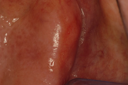 [Figure caption and citation for the preceding image starts]: 红斑承蒙 James Sciubba 医生提供;准许使用 [Citation ends].至少 85% 的病例病理表现为癌或重度上皮异常增生。白斑也属于潜在恶性病损,尤其是伴有红色病损的白斑(颗粒状白斑)。
 [Figure caption and citation for the preceding image starts]: 颗粒状白斑承蒙 James Sciubba 医生提供;准许使用 [Citation ends].利用二元显微分类系统,可将病变分为低风险异常增生或高风险异常增生,后者是评估恶性转变的重要指标。[58]Liu W, Wang YF, Zhou HW, et al. Malignant transformation of oral leukoplakia: a retrospective cohort study of 218 Chinese patients. BMC Cancer. 2010;10:685.http://www.ncbi.nlm.nih.gov/pmc/articles/PMC3009685/?tool=pubmedhttp://www.ncbi.nlm.nih.gov/pubmed/21159209?tool=bestpractice.com包括增殖性疣状白斑、
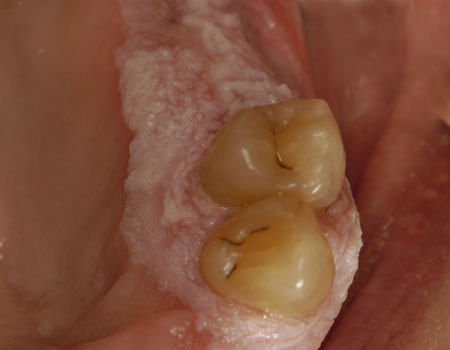 [Figure caption and citation for the preceding image starts]: 增殖性疣状白斑承蒙 James Sciubba 医生提供;准许使用 [Citation ends].舌下白斑(舌下角化症)、
 [Figure caption and citation for the preceding image starts]: 舌下白斑承蒙 James Sciubba 医生提供;准许使用 [Citation ends].和念珠菌性白斑
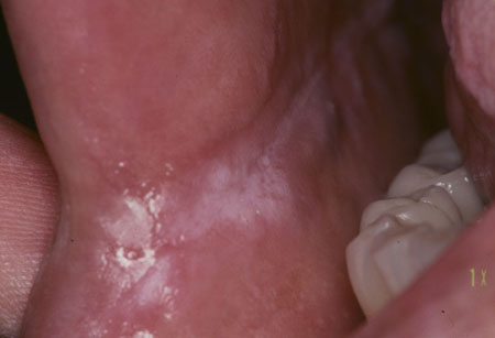 [Figure caption and citation for the preceding image starts]: 念珠菌白斑承蒙 James Sciubba 医生提供;准许使用 [Citation ends].在内的病损也具有恶变潜能。和其他类型的白斑相比,增殖性疣状白斑癌变几率更高。对美国≥65 岁成人开展的一项大型病例队列研究发现,相对于口腔鳞状细胞癌但无既往粘膜白斑病的患者,既往患有粘膜白斑病的口腔癌患者,其癌症相关的死亡风险更低。研究得出结论,粘膜白斑病的识别与分析有助于早期癌症检测,改善生存水平。[59]Yanik EL, Katki HA, Silverberg MJ, et al. Leukoplakia, oral cavity cancer risk, and cancer survival in the US elderly. Cancer Prev Res (Phila). 2015;8:857-863.http://www.ncbi.nlm.nih.gov/pubmed/4560597?tool=bestpractice.com
体格检查
必须进行彻底的口腔和局部淋巴结检查。很多潜在恶性口腔病损(包括白斑)和癌可以通过视诊被查出,但在检查不彻底的情况下很容易被忽略掉。[51]Sankaranarayanan R, Fernandez Garrote L, Lence Anta J, et al. Visual inspection in oral cancer screening in Cuba: a case-control study. Oral Oncol. 2002;38:131-136.http://www.ncbi.nlm.nih.gov/pubmed/11854059?tool=bestpractice.com[52]Shugars DC, Patton LL. Detecting, diagnosing, and preventing oral cancer. Nurse Pract 1997;22:105, 109-110, 113-115.http://www.ncbi.nlm.nih.gov/pubmed/9211456?tool=bestpractice.com[60]Massano J, Regateiro FS, Januario G, et al. Oral squamous cell carcinoma: review of prognostic and predictive factors. Oral Surg Oral Med Oral Pathol Oral Radiol Endod. 2006;102:67-76.http://www.ncbi.nlm.nih.gov/pubmed/16831675?tool=bestpractice.com若口内出现广泛的黏膜病损(“区域改变”)或第二肿瘤,则提示应该对口腔黏膜进行全面彻底的检查。[61]Slaughter DP, Southwick HW, Smejkal W. Field cancerization in oral stratified squamous epithelium; clinical implications of multicentric origin. Cancer. 1953;6;963-968.http://www.ncbi.nlm.nih.gov/pubmed/13094644?tool=bestpractice.com分子水平的变化可提示恶变潜能,但临床上可能并没有相应的明显的病损,甚至可能没有可以分辨的病损。[62]Braakhuis BJ, Brakenhoff RH, Leemans CR. Head and neck cancer: molecular carcinogenesis. Ann Oncol. 2005;16(suppl 2):ii249-ii250.http://annonc.oxfordjournals.org/cgi/reprint/16/suppl_2/ii249http://www.ncbi.nlm.nih.gov/pubmed/15958466?tool=bestpractice.com[63]Braakhuis BJ, Leemans CR, Brakenhoff RH. Expanding fields of genetically altered cells in head and neck squamous carcinogenesis. Semin Cancer Biol. 2005;15:113-120.http://www.ncbi.nlm.nih.gov/pubmed/15652456?tool=bestpractice.com[64]Tabor MP, Brakenhoff RH, Ruijter-Schippers HJ, et al. Genetically altered fields as origin of locally recurrent head and neck cancer: a retrospective study. Clin Cancer Res. 2004;10:3607-3613.http://clincancerres.aacrjournals.org/content/10/11/3607.fullhttp://www.ncbi.nlm.nih.gov/pubmed/15173066?tool=bestpractice.com[65]Braakhuis BJ, Tabor MP, Kummer JA, et al. A genetic explanation of Slaughter's concept of field cancerization: evidence and clinical implications. Cancer Res. 2003;63:1727-1730.http://cancerres.aacrjournals.org/cgi/content/full/63/8/1727http://www.ncbi.nlm.nih.gov/pubmed/12702551?tool=bestpractice.com这也部分解释了口腔癌前病变和口腔癌的患者出现第二肿瘤原发灶的风险增高的原因。[66]Lippman SM, Hong WK. Second malignant tumors in head and neck squamous cell carcinoma: the overshadowing threat for patients with early-stage disease. Int J Radiat Oncol Biol Phys. 1989;17:691-694.http://www.ncbi.nlm.nih.gov/pubmed/2674081?tool=bestpractice.com
某些常见疾病也会给诊断带来麻烦,包括福代斯斑和地图舌。口内积聚的食物残渣(软垢)或真菌(念珠菌病)也可表现为白色斑片,但是通常很容易被纱球拭去。其他病损呈白色外观,通常是上皮角质层增厚所致。 [Figure caption and citation for the preceding image starts]: 均质型白斑承蒙 James Sciubba 医生提供;准许使用 [Citation ends].口腔毛状白斑是由病毒引起的病损。主要发生在免疫抑制(例如 HIV 感染)的人群中,其恶变潜能并不明确。
 [Figure caption and citation for the preceding image starts]: 口腔毛状白斑承蒙 James Sciubba 医生提供;准许使用 [Citation ends].
白色病损通常是无痛的,可为局灶性、多灶性、条纹状或弥散性分布,这些特征对诊断有指导作用。例如:
咬合线区域的局限性病损常常由咬颊引起,表现为塌陷的疱样病损或白角化 [Figure caption and citation for the preceding image starts]: 咬唇-颊症承蒙 James Sciubba 医生提供;准许使用 [Citation ends].
多灶性病损在鹅口疮(假膜型念珠菌病)和扁平苔癣患者中比较常见;条纹状病损是扁平苔癣的典型症状
弥散性白色病损可见于白水肿的颊黏膜和烟碱性口炎的腭黏膜。
必须排除引起口腔白色病损的其他病因。在排除了这些因素后,即可诊断为白斑。
口腔白色病损的病因
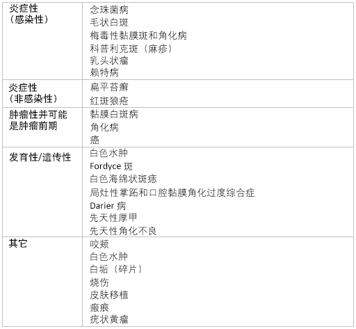 [Figure caption and citation for the preceding image starts]: 口腔白色病损的病因由 BMJ 证据中心团队创建 [Citation ends].
排除其他诊断的相关检测
多数情况下通过详尽的病史采集和体格检查即可做出诊断,但在某些情况下,只有对代表性病损区域进行活检后才能确诊。额外的实验室检查可能有助于排除诊断。其中可这些检查包括梅毒螺旋体血清学测定和自体抗体检测。
一线检测
白斑的恶变潜能很难判断,这需要对特征性临床表现及其转归进行准确的评估。然而,临床诊断本身并不足以确定和排除恶性和潜在恶性病损;即使是经验丰富、且受过良好培训的专业人员也不能单凭视诊就确诊所有癌前病变和早期 OSCC。[67]Silverman S Jr. Early diagnosis of oral cancer. Cancer. 1988;62(suppl 8):1796-1799.http://www.ncbi.nlm.nih.gov/pubmed/3167796?tool=bestpractice.com此外,公认的 OSCC 的典型特征,例如溃疡、硬结、膨隆、出血和颈淋巴结肿大,都是疾病的晚期症状,而并非早期症状。[68]Mashberg A, Feldman LJ. Clinical criteria for identifying early oral and oropharyngeal carcinoma: erythroplasia revisited. Am J Surg. 1988;156:273-275.http://www.ncbi.nlm.nih.gov/pubmed/3177749?tool=bestpractice.com遗憾的是,临床数据表明,即使口腔病损已经明显表现为癌,还是经常发生活检的延误。[69]Scully C, Malamos D, Levers BG, et al. Sources and patterns of referrals of oral cancer: role of general practitioners. Br Med J (Clin Res Ed). 1986;293:599-601.http://www.pubmedcentral.nih.gov/articlerender.fcgi?artid=1341389http://www.ncbi.nlm.nih.gov/pubmed/3092946?tool=bestpractice.com[70]Dimitroulis G, Reade P, Wiesenfeld D. Referral patterns of patients with oral squamous cell carcinoma, Australia. Eur J Cancer B Oral Oncol. 1992;28B:23-27.http://www.ncbi.nlm.nih.gov/pubmed/1422466?tool=bestpractice.com
活检
白斑切取活检适用于大多数白斑,切取时应选取具有代表性的病损区域,切取的组织要足够大。应避免进行切除活检,因为如果病损证实为恶性,切除病损时不太可能切至合适的组织边界。而且这也可能影响外科医生对病损部位和特征的准确判断。特征。
研究表明,切取活检偶尔会出现假阴性结果。研究发现,将已通过切取活检排除了异常增生的白斑病损再进行切除活检,其中高达 10% 的病损可查出 OSCC 病灶。[71]Chiesa F, Sala L, Costa L, et al. Excision of oral leukoplakias by CO2 laser on an outpatient basis: a useful procedure for prevention and early detection of oral carcinomas. Tumori. 1986;72:307-312.http://www.ncbi.nlm.nih.gov/pubmed/3739009?tool=bestpractice.com[72]Holmstrup P, Vedtofte P, Reibel J, et al. Oral premalignant lesions: is a biopsy reliable? J Oral Pathol Med. 2007;36:262-266.http://www.ncbi.nlm.nih.gov/pubmed/17448135?tool=bestpractice.com数据表明,早期恶性病损可发生在潜在恶性病损的区域,甚至超出该范围,这在一定程度上解释了上述现象。[63]Braakhuis BJ, Leemans CR, Brakenhoff RH. Expanding fields of genetically altered cells in head and neck squamous carcinogenesis. Semin Cancer Biol. 2005;15:113-120.http://www.ncbi.nlm.nih.gov/pubmed/15652456?tool=bestpractice.com[73]Califano J, Westra WH, Meininger G, et al. Genetic progression and clonal relationship of recurrent premalignant head and neck lesions. Clin Cancer Res. 2000;6:347-352.http://clincancerres.aacrjournals.org/cgi/content/full/6/2/347http://www.ncbi.nlm.nih.gov/pubmed/10690509?tool=bestpractice.com活检时应当选择病损中的红色区域,因为红色区域比白色区域更可能出现异常增生。
病理学
由经验丰富的病理学家来进行组织病理学分析对于治疗至关重要,因为病理学家之间和病理医生本身的差异性不容忽视。[74]Abbey LM, Kaugars GE, Gunsolley JC, et al. Intraexaminer and interexaminer reliability in the diagnosis of oral epithelial dysplasia. Oral Surg Oral Med Oral Pathol Oral Radiol Endod. 1995;80:188-191.http://www.ncbi.nlm.nih.gov/pubmed/7552884?tool=bestpractice.com[75]Karabulut A, Reibel J, Therkildsen MH, et al. Observer variability in the histologic assessment of oral premalignant lesions. J Oral Pathol Med. 1995;24:198-200.http://www.ncbi.nlm.nih.gov/pubmed/7616457?tool=bestpractice.com[76]Fischer DJ, Epstein JB, Morton TH, et al. Interobserver reliability in the histopathologic diagnosis of oral premalignant and malignant lesions. J Oral Pathol Med. 2004;33:65-70.http://www.ncbi.nlm.nih.gov/pubmed/14720191?tool=bestpractice.com[77]Fischer DJ, Epstein JB, Morton TH Jr, et al. Reliability of histologic diagnosis of clinically normal intraoral tissue adjacent to clinically suspicious lesions in former upper aerodigestive tract cancer patients. Oral Oncol. 2005;41:489-496.http://www.ncbi.nlm.nih.gov/pubmed/15878753?tool=bestpractice.com[78]Woolgar JA. Histopathological prognosticators in oral and oropharyngeal squamous cell carcinoma. Oral Oncol. 2006;42:229-239.http://www.ncbi.nlm.nih.gov/pubmed/16150633?tool=bestpractice.com如果在初次活检后,临床医生对活检部位的代表性存有疑问(例如,病理报告显示无恶性病损,但是临床上怀疑存在恶性病损),可行二次活检。
常规组织学分析表明,白斑具有多样化的组织学特性,从良性到不同程度的异常增生。良性的组织学特征为正角化/角化不全,且上皮深至表层的区域内无角蛋白形成(角化不良是发育异常的表现之一)。此外,通常会出现棘细胞层增厚或总体积增大(棘层增厚)。值得注意的是,大多数白斑(超过 60%)镜下仅表现为过角化伴或不伴棘层增厚。上皮基底层和更表浅的细胞层常无明确的细胞异型性。然而,最近在泰国人群中的一项研究发现,10.6% 的白斑实际上伴有异常增生,且其中 4.9% 被诊断为鳞状细胞癌。[79]Lathanasupkul P, Poomsawat S, Punyasingh J. A clinicopathologic study of oral leukoplakia and erythroplakia in a Thai population. Quintessence Int. 2007;38:e448-e455.http://www.ncbi.nlm.nih.gov/pubmed/17823667?tool=bestpractice.com
当存在上皮异常增生时,应该对其评估进行分级(例如轻度、中度、重度);此外,上皮内瘤变(这一概念最早用于子宫颈黏膜,后来扩展到其他黏膜)也被一些学者应用于口腔黏膜病损的诊断(口腔上皮内瘤变)。[80]Kuffer R, Lombardi T. Premalignant lesions of the oral mucosa: a discussion about the place of oral intraepithelial neoplasia (OIN). Oral Oncol. 2002;38:125-130.http://www.ncbi.nlm.nih.gov/pubmed/11854058?tool=bestpractice.com与癌前病变或上皮异常增生相关的病理变化如下:
核多形性
极性消失
有丝分裂象增加
核质比变化
角化不良,或上皮深至表面的区域内有角蛋白存在
上皮层次紊乱
细胞间黏附力下降或细胞附着结构减少
辅助诊断技术
现有多种辅助诊断技术可用于口腔黏膜病理的临床评估。但这些技术的有效性尚缺乏足够证据支持。[81]Rethman M, Carpenter W, Cohen EE, et al. Evidence-based clinical recommendations regarding screening for oral squamous cell carcinomas. J Am Dent Assoc. 2010;14:509-520.http://www.ncbi.nlm.nih.gov/pubmed/20436098?tool=bestpractice.com[82]Fedele S. Diagnostic aids in the screening of oral cancer. Head Neck Oncol. 2009;1:5.http://www.ncbi.nlm.nih.gov/pmc/articles/PMC2654034/?tool=pubmedhttp://www.ncbi.nlm.nih.gov/pubmed/19284694?tool=bestpractice.com具体技术包括:
毛刷活检
对于那些没有足够切取活检指征的、普通的、表观无害的口腔病损,可以使用口腔毛刷活检来检测是否存在上皮异常增生。这一检测使用小尼龙刷子收集病损区域的细胞样品,然后对样品进行电脑分析。样本采集简单,不引起或仅引起轻微疼痛或出血,无需麻醉;但是,必须对病损部位进行准确采样。细胞检测的结果和病理医生的书面报告将被反馈给临床医生,如果检测发现显著异常,将建议行常规切取活检。
据报道,在上皮异常增生和 OSCC 检测中,这一检查的灵敏度高于 92%至 96%,特异性高于 90%至 94%,[83]Sciubba JJ. Improving detection of precancerous and cancerous oral lesions: computer-assisted analysis of the oral brush biopsy. U.S. Collaborative OralCDx Study Group. J Am Dent Assoc. 1999;130:1445-1457.http://www.ncbi.nlm.nih.gov/pubmed/10570588?tool=bestpractice.com[84]Scheifele C, Schmidt-Westhausen A, Dietrich T, et al. The sensitivity and specificity of the OralCDx technique: evaluation of 103 cases. Oral Oncol. 2004;40:824-828.http://www.ncbi.nlm.nih.gov/pubmed/15288838?tool=bestpractice.com阳性预测值在 38%至 44%之间。[85]Svirsky JA, Burns JC, Carpenter WM, et al. Comparison of computer-assisted brush biopsy results with follow up scalpel biopsy and histology. Gen Dent. 2002;50:500-503.http://www.ncbi.nlm.nih.gov/pubmed/12572180?tool=bestpractice.com[86]Kosicki DM, Riva C, Pajarola GF, et al. Oral CDx brush biopsy - a tool for early diagnosis of oral squamous cell carcinoma [in German]. Schweiz Monatsschr Zahnmed. 2007;117:222-227.http://www.ncbi.nlm.nih.gov/pubmed/17425240?tool=bestpractice.com[87]Poate TW, Buchanan JA, Hodgson TA, et al. An audit of the efficacy of the oral brush biopsy technique in a specialist Oral Medicine unit. Oral Oncol. 2004;40:829-834.http://www.ncbi.nlm.nih.gov/pubmed/15288839?tool=bestpractice.com毛刷活检现已被用于检测在切取活检中遗漏的 OSCC。[88]Remmerbach TW, Weidenbach H, Hemprich A, et al. Earliest detection of oral cancer using non-invasive brush biopsy including DNA-image-cytometry: report on four cases. Anal Cell Pathol. 2003;25:159-166.http://www.ncbi.nlm.nih.gov/pubmed/14501082?tool=bestpractice.com但是,该检测不能检测出全部潜在恶性病变。部分 OSCC 患者的毛刷活检结果可为阴性,目前已有这方面的病案报告。[89]Potter TJ, Summerlin DJ, Campbell JH. Oral malignancies associated with negative transepithelial brush biopsy. J Oral Maxillofac Surg. 2003;61:674-677.http://www.ncbi.nlm.nih.gov/pubmed/12796875?tool=bestpractice.com
使用分子技术(例如 p53 突变、DNA 定量分析和 AgNOR(通过银染技术使核仁形成区蛋白计数可视化)可以改善毛刷活检结果。[90]Scheifele C, Schlechte H, Bethke G, et al. Detection of TP53-mutations in brush biopsies from oral leukoplakias [in German]. Mund Kiefer Gesichtschir. 2002;6:410-414.http://www.ncbi.nlm.nih.gov/pubmed/12447653?tool=bestpractice.com[91]Remmerbach TW, Mathes SN, Weidenbach H, et al. Noninvasive brush biopsy as an innovative tool for early detection of oral carcinomas [in German]. Mund Kiefer Gesichtschir. 2004;8:229-236.http://www.ncbi.nlm.nih.gov/pubmed/15293118?tool=bestpractice.com[92]Maraki D, Becker J, Boecking A. Cytologic and DNA-cytometric very early diagnosis of oral cancer. J Oral Pathol Med. 2004;33:398-404.http://www.ncbi.nlm.nih.gov/pubmed/15250831?tool=bestpractice.com[93]Remmerbach TW, Weidenbach H, Muller C, et al. Diagnostic value of nucleolar organizer regions (AgNORs) in brush biopsies of suspicious lesions of the oral cavity. Anal Cell Pathol. 2003;25:139-146.http://www.ncbi.nlm.nih.gov/pubmed/12775918?tool=bestpractice.com
分子和染色体标志物
在对癌前病变进行临床和病理分析的基础上,想要实现对其恶性转归的预测,关键是要发掘使用简便、经济实用的分子标志物。在对患者进行的常规随访中,这些技术是十分有用的。
近期研究发现了数个在预测病损发展趋势方面具有潜在价值的分子标志物。[15]Zhang L, Rosin MP. Loss of heterozygosity: a potential tool in management of oral premalignant lesions? J Oral Pathol Med. 2001;30:513-520.http://www.ncbi.nlm.nih.gov/pubmed/11555152?tool=bestpractice.com例如,在对发生于危险区域的白斑进行的评估中发现,遗传变异水平的提高与癌变风险的增加相关,如染色体 3p 和/或 9p 位点杂合性缺失。[94]Zhang L, Cheung KJ Jr, Lam WL, et al. Increased genetic damage in oral leukoplakia from high risk sites: potential impact on staging and clinical management. Cancer. 2001;91:2148-2155.http://www3.interscience.wiley.com/cgi-bin/fulltext/82003264/HTMLSTARThttp://www.ncbi.nlm.nih.gov/pubmed/11391596?tool=bestpractice.com平足蛋白是一种跨膜糖蛋白,可能成为将来对口腔白斑患者的恶性转化风险进行评估的重要生化标志物。[95]de Vicente JC, Rodrigo JP, Rodriguez-Santamarta T, et al. Podoplanin expression in oral leukoplakia: tumorigenic role. Oral Oncol. 2013;49:598-603.http://www.ncbi.nlm.nih.gov/pubmed/23473850?tool=bestpractice.com此外,基因芯片分析技术和其他分子诊断工具在白斑癌变的预测方面也具有应用前景。[96]Patel V, Leethanakul C, Gutkind JS. New approaches to the understanding of the molecular basis of oral cancer. Crit Rev Oral Biol Med. 2001;12:55-63.http://www.ncbi.nlm.nih.gov/pubmed/11349962?tool=bestpractice.com
在评估口腔白斑时,DNA 倍体状态仍是值得关注的一个方面,已有研究显示非整倍体与口腔白斑癌变风险的增加相关。[97]Bremmer JF, Brakenhoff RH, Broeckaert MA, et al. Prognostic value of DNA ploidy status in patients with oral leukoplakia. Oral Oncol. 2011;47:956-960.http://www.ncbi.nlm.nih.gov/pubmed/21880540?tool=bestpractice.com此外,伴上皮异常增生的白斑病损的 S 期分数显示其非整倍体的比率是不伴上皮异常增生的白斑的两倍以上。[98]Khanna R, Agarwal A, Khanna S, et al. S-phase fraction and DNA ploidy in oral leukoplakia. ANZ J Surg. 2010;80:548-551.http://www.ncbi.nlm.nih.gov/pubmed/20795971?tool=bestpractice.com研究者利用逆转录酶 PCR 进行微阵列分析,在发生癌变的白斑病例中发现了一些候选分子标志物,进一步加深了我们对口腔白斑癌变分子机制的理解。[99]Liu W, Zheng W, Xie J, et al. Identification of genes related to carcinogenesis of oral leukoplakia by oligo cancer microarray analysis. Oncol Rep. 2011;26:265-274.http://www.ncbi.nlm.nih.gov/pubmed/21523324?tool=bestpractice.com
光学系统
光谱学系统可通过实时、无创的方式进行组织诊断,其潜在应用价值越来越受到人们的关注。[100]Swinson B, Jerjes W, El-Maaytah M, et al. Optical techniques in diagnosis of head and neck malignancy. Oral Oncol. 2006;42:221-228.http://www.ncbi.nlm.nih.gov/pubmed/16140566?tool=bestpractice.com[101]Suhr MA, Hopper C, Jones L, et al. Optical biopsy systems for the diagnosis and monitoring of superficial cancer and precancer. Int J Oral Maxillofac Surg. 2000;29:453-457.http://www.ncbi.nlm.nih.gov/pubmed/11202330?tool=bestpractice.com[102]Sharwani A, Jerjes W, Salih V, et al. Assessment of oral premalignancy using elastic scattering spectroscopy. Oral Oncol. 2006;42:343-349.http://www.ncbi.nlm.nih.gov/pubmed/16321565?tool=bestpractice.com在实际操作时,先用醋酸溶液漱口,然后使用化学发光法对黏膜进行检查。这类光学辅助手段有助于识别黏膜病损,选择活检部位。同时可以帮助识别肉眼常规检查无法发现的细胞和分子异常,并能提供病损旁组织的相关信息。[103]Westra WH, Sidransky D. Fluorescence visualization in oral neoplasia: shedding light on an old problem. Clin Cancer Res. 2006;12:6594-6597.http://clincancerres.aacrjournals.org/cgi/content/full/12/22/6594http://www.ncbi.nlm.nih.gov/pubmed/17121876?tool=bestpractice.com[104]Rosin MP, Cheng X, Poh C, et al. Use of allelic loss to predict malignant risk for low-grade oral epithelial dysplasia. Clin Cancer Res. 2000;6:357-362.http://clincancerres.aacrjournals.org/cgi/content/full/6/2/357http://www.ncbi.nlm.nih.gov/pubmed/10690511?tool=bestpractice.com
然而,关于这类技术的临床获益的研究数据并不一致;研究发现,这一技术对上皮异常增生病损的检出率与传统临床诊断以及活检部位选择的结果类似。[105]Oh ES, Laskin DM. Efficacy of the ViziLite system in the identification of oral lesions. J Oral Maxillofac Surg. 2007;65:424-426.http://www.ncbi.nlm.nih.gov/pubmed/17307587?tool=bestpractice.com[106]Epstein JB, Gorsky M, Lonky S, et al. The efficacy of oral lumenoscopy (ViziLite) in visualizing oral mucosal lesions. Spec Care Dentist. 2006;26:171-174.http://www.ncbi.nlm.nih.gov/pubmed/16927741?tool=bestpractice.com[107]Lane PM, Gilhuly T, Whitehead P, et al. Simple device for the direct visualization of oral-cavity tissue fluorescence. J Biomed Opt. 2006;11:024006.http://www.ncbi.nlm.nih.gov/pubmed/16674196?tool=bestpractice.com[108]Poh CF, Ng SP, Williams PM, et al. Direct fluorescence visualization of clinically occult high-risk oral premalignant disease using a simple hand-held device. Head Neck. 2007;29:71-76.http://www.ncbi.nlm.nih.gov/pubmed/16983693?tool=bestpractice.com[109]Poh CF, Zhang L, Anderson DW, et al. Fluorescence visualization detection of field alterations in tumor margins of oral cancer patients. Clin Cancer Res. 2006;12:6716-6722.http://clincancerres.aacrjournals.org/cgi/content/full/12/22/6716http://www.ncbi.nlm.nih.gov/pubmed/17121891?tool=bestpractice.com同时,采用这些光学辅助手段检测到的口腔病损,其病理学改变仍需常规活检来证实。
