针对青少年特发性脊柱侧凸 (AIS) 的治疗决策基于就诊时的初始畸形、侧凸进展速度,以及患者生长潜能的整体评估。
治疗的主要目标是防止脊柱畸形进展,直到患者骨骼发育成熟。一旦患者骨骼发育成熟,侧凸进展风险就显著下降。AIS 治疗选择包括观察性监测、支具固定或脊柱关节固定术(融合)。根据侧凸的严重程度以及直到骨骼发育成熟的剩余生长量选择治疗。
筛查时脊柱侧凸测量仪测量值<5° 或站立位冠状面 Cobb 角测量值≤10°
依据这些测量值,可诊断为姿势性畸形。无需治疗或观察性监测,除非患者或其家属发现患者姿势不对称的情况恶化,或者患者出现提示脊柱畸形其他潜在原因的症状。[2]Newton PO, Wenger DR. Idiopathic scoliosis. In Morrissy RT, Weinstein SL, eds. Lovell and Winter's pediatric orthopaedics. 6th ed. Philadelphia: Lippincott, Williams & Wilkins; 2006:693-762.[12]Bunnell WP. The natural history of idiopathic scoliosis before skeletal maturity. Spine. 1986;11:773-776.[1]Parent S, Newton PO, Wenger DR. Adolescent idiopathic scoliosis: etiology, anatomy, natural history, and bracing. Instr Course Lect. 2005;54:529-536.[40]Weinstein SL, Dolan LA, Spratt KF, et al. Health and function of patients with untreated idiopathic scoliosis: a 50-year natural history study. JAMA. 2003;289:559-567.http://jama.ama-assn.org/cgi/content/full/289/5/559[41]Bagnall KM, Beuerlein M, Johnson P, et al. Pineal transplantation after pinealectomy in young chickens has no effect on the development of scoliosis. Spine. 2001;26:1022-1027.[42]Goldberg MS, Mayo NE, Poitras B, et al. The Ste-Justine Adolescent Idiopathic Scoliosis Cohort Study. Part II: Perception of health, self and body image, and participation in physical activities. Spine. 1994;19:1562-1572.[43]Weinstein SL. Idiopathic scoliosis. Natural history. Spine. 1986;11:780-783.[44]Weinstein SL, Ponseti IV. Curve progression in idiopathic scoliosis. J Bone Joint Surg Am. 1983;65:447-455.[45]Weinstein SL, Zavala DC, Ponseti IV. Idiopathic scoliosis: long-term follow-up and prognosis in untreated patients. J Bone Joint Surg Am. 1981;63:702-712.
应建议患者维持规律的锻炼计划,尤其应加强中枢肌肉力量和健身训练。
站立位冠状面 Cobb 角测量值为 11°-20°
应进行观察性监测,每 4 到 12 个月(具体间隔取决于评估时的生长速度和剩余生长潜能)随访一次站立时后前位和侧位脊柱侧凸 X 线片。[2]Newton PO, Wenger DR. Idiopathic scoliosis. In Morrissy RT, Weinstein SL, eds. Lovell and Winter's pediatric orthopaedics. 6th ed. Philadelphia: Lippincott, Williams & Wilkins; 2006:693-762.[12]Bunnell WP. The natural history of idiopathic scoliosis before skeletal maturity. Spine. 1986;11:773-776.[1]Parent S, Newton PO, Wenger DR. Adolescent idiopathic scoliosis: etiology, anatomy, natural history, and bracing. Instr Course Lect. 2005;54:529-536.[40]Weinstein SL, Dolan LA, Spratt KF, et al. Health and function of patients with untreated idiopathic scoliosis: a 50-year natural history study. JAMA. 2003;289:559-567.http://jama.ama-assn.org/cgi/content/full/289/5/559[41]Bagnall KM, Beuerlein M, Johnson P, et al. Pineal transplantation after pinealectomy in young chickens has no effect on the development of scoliosis. Spine. 2001;26:1022-1027.[42]Goldberg MS, Mayo NE, Poitras B, et al. The Ste-Justine Adolescent Idiopathic Scoliosis Cohort Study. Part II: Perception of health, self and body image, and participation in physical activities. Spine. 1994;19:1562-1572.[43]Weinstein SL. Idiopathic scoliosis. Natural history. Spine. 1986;11:780-783.[44]Weinstein SL, Ponseti IV. Curve progression in idiopathic scoliosis. J Bone Joint Surg Am. 1983;65:447-455.[45]Weinstein SL, Zavala DC, Ponseti IV. Idiopathic scoliosis: long-term follow-up and prognosis in untreated patients. J Bone Joint Surg Am. 1981;63:702-712.
应建议患者维持规律的锻炼计划,尤其应加强中枢肌肉力量和健身训练。
站立位冠状面 Cobb 角测量值为 21°-45°
传统上,患者侧凸>21° 时采用支具治疗。[2]Newton PO, Wenger DR. Idiopathic scoliosis. In Morrissy RT, Weinstein SL, eds. Lovell and Winter's pediatric orthopaedics. 6th ed. Philadelphia: Lippincott, Williams & Wilkins; 2006:693-762.[1]Parent S, Newton PO, Wenger DR. Adolescent idiopathic scoliosis: etiology, anatomy, natural history, and bracing. Instr Course Lect. 2005;54:529-536. 有多种支具可供选择,所用的具体矫形器具类型因外科医师和国家/地区而异。[46]Sponseller PD. Bracing for adolescent idiopathic scoliosis in practice today. J Pediatr Orthop. 2011;31:S53-S60. 美国使用的支具通常是 Boston 支具、Rosenberger 支具和 Charleston 屈曲支具。
从每天 23 小时穿戴支具到仅夜间穿戴支具,多种支具穿戴计划也可供考虑。虽然一些研究认为支具穿戴与侧凸进展之间存在剂量依赖性关系,但仍不明确哪种支具穿戴计划带来的结局最为可靠。脊柱侧凸研究学会共识支持每天穿戴 18 小时的支具计划。Scoliosis Research Society 其他组织同样尝试制定“治疗共识”声明和指南,以试图规范脊柱侧凸支具治疗方案。[3]Negrini S, Grivas TB, Kotwicki T, et al; International Society on Scoliosis Orthopaedic and Rehabilitation Treatment (SOSORT). Guidelines on "Standards of management of idiopathic scoliosis with corrective braces in everyday clinics and in clinical research": SOSORT Consensus 2008. Scoliosis. 2009;4:2.http://www.scoliosisjournal.com/content/4/1/2[47]Kotwicki T, Durmala J, Czaprowski D, et al. Conservative management of idiopathic scoliosis: guidelines based on SOSORT 2006 Consensus. Ortop Traumatol Rehabil. 2009;11:379-395.
支具治疗的目的是防止侧凸进展,直到患者骨骼发育成熟,届时侧凸进一步进展的风险会显著下降。来自青少年特发性脊柱侧凸支具试验 (BrAIST) 的数据显示,与未穿戴支具的患者相比,穿戴支具的 AIS 患者发生侧凸进展的风险明显降低。[48]Dolan LA, Wright JG, Weinstein SL. Effects of bracing in adolescents with idiopathic scoliosis. N Engl J Med. 2014;370:681.http://www.nejm.org/doi/full/10.1056/NEJMc1314229 一项 Cochrane 评价总结称,所有纳入研究(包括 7 项共涉及 662 名参与者的单独研究)一致显示 AIS 患者穿戴支具防止了侧凸进展。[49]Negrini S, Minozzi S, Bettany-Saltikov J, et al. Braces for idiopathic scoliosis in adolescents. Cochrane Database Syst Rev. 2015;(6):CD006850.http://onlinelibrary.wiley.com/doi/10.1002/14651858.CD006850.pub3/full
应建议患者维持规律的锻炼计划,尤其应加强中枢肌肉力量和健身训练。 [Figure caption and citation for the preceding image starts]: 典型的胸腰骶矫形器 (TLSO) ——脊柱侧凸支具Weinstein SL, et al. Adolescent idiopathic scoliosis.Lancet.2008;371:1527-1537. 经授权使用 [Citation ends].
站立位冠状面 Cobb 角测量值为>45°
由于侧凸进一步进展的风险以及成年时相关的并发症,对于此组患者,建议的治疗为进行脊柱关节固定术。脊柱关节融合内固定术具有多项治疗目标,包括矫正主弯、通过躯干平衡改善形体、防止侧凸继续进展,以及减少与脊柱畸形有关的短期和长期并发症。[50]Dickson JH, Mirkovic S, Noble PC, et al. Results of operative treatment of idiopathic scoliosis in adults. J Bone Joint Surg Am. 1995;77:513-523.
选择哪种手术方法和器械技术取决于畸形的特征(例如侧凸顶椎的位置)、脊柱柔韧度和外科医生的偏好。[50]Dickson JH, Mirkovic S, Noble PC, et al. Results of operative treatment of idiopathic scoliosis in adults. J Bone Joint Surg Am. 1995;77:513-523. 大部分畸形都能通过后入路进行矫正,传统上它是脊柱侧凸关节融合术的最优技术。[51]Kim YJ, Lenke LG, Kim J, et al. Comparative analysis of pedicle screw versus hybrid instrumentation in posterior spinal fusion of adolescent idiopathic scoliosis. Spine. 2006;31:291-298.[52]Lenke LG. Debate: Resolved, a 55 degrees right thoracic adolescent idiopathic scoliotic curve should be treated by posterior spinal fusion and segmental instrumentation using thoracic pedicle screws: Pro: Thoracic pedicle screws should be used to treat a 55 degrees right thoracic adolescent idiopathic scoliosis. J Pediatr Orthop. 2004;24:329-234.[53]Suk SI, Lee CK, Kim WJ, et al. Segmental pedicle screw fixation in the treatment of thoracic idiopathic scoliosis. Spine. 1995;20:1399-1405.[54]Suk SI, Lee SM, Chung ER, et al. Selective thoracic fusion with segmental pedicle screw fixation in the treatment of thoracic idiopathic scoliosis: more than 5-year follow-up. Spine. 2005;30:1602-1609.一些外科医师采用前入路,他们认为可通过更少的融合节段矫正畸形。[55]Muschik MT, Kimmich H, Demmel T. Comparison of anterior and posterior double-rod instrumentation for thoracic idiopathic scoliosis: results of 141 patients. Eur Spine J. 2006;15:1128-1138.[56]Turi M, Johnston CE 2nd, Richards BS. Anterior correction of idiopathic scoliosis using TSRH instrumentation. Spine. 1993;18:417-422.[57]Newton PO. The use of video-assisted thoracoscopic surgery in the treatment of adolescent idiopathic scoliosis. Instr Course Lect. 2005;54:551-558.[58]Newton PO, Faro FD, Gollogly S, et al. Results of preoperative pulmonary function testing of adolescents with idiopathic scoliosis. A study of six hundred and thirty-one patients. J Bone Joint Surg Am. 2005;87:1937-1946.[59]Newton PO, Marks M, Faro F, et al. Use of video-assisted thoracoscopic surgery to reduce perioperative morbidity in scoliosis surgery. Spine. 2003;28:S249-S254.[60]Newton PO, Parent S, Marks M, et al. Prospective evaluation of 50 consecutive scoliosis patients surgically treated with thoracoscopic anterior instrumentation. Spine. 2005;30:S100-S109.[61]Newton PO, Wenger DR, Mubarak SJ, et al. Anterior release and fusion in pediatric spinal deformity. A comparison of early outcome and cost of thoracoscopic and open thoracotomy approaches. Spine. 1997;22:1398-1406.[62]Newton PO, White KK, Faro F, et al. The success of thoracoscopic anterior fusion in a consecutive series of 112 pediatric spinal deformity cases. Spine. 2005;30:392-398.[63]Reddi V, Clarke DV Jr, Arlet V. Anterior thoracoscopic instrumentation in adolescent idiopathic scoliosis: a systematic review. Spine (Phila Pa 1976). 2008;33:1986-1994. 然而,前入路手术的植入失败和假关节发生率更高,而且由于手术期间需要“单肺”麻醉,因此有引起肺部并发症的风险。[64]Betz RR, Harms J, Clements DH 3rd, et al. Comparison of anterior and posterior instrumentation for correction of adolescent thoracic idiopathic scoliosis. Spine. 1999;24:225-239.[65]Lenke LG. Anterior endoscopic discectomy and fusion for adolescent idiopathic scoliosis. Spine. 2003;28:S36-S43.[66]Lowe TG, Alongi PR, Smith DA, et al. Anterior single rod instrumentation for thoracolumbar adolescent idiopathic scoliosis with and without the use of structural interbody support. Spine. 2003;28:2232-2241.
应建议患者维持规律的锻炼计划,尤其应加强中枢肌肉力量和健身训练。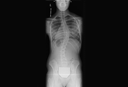 [Figure caption and citation for the preceding image starts]: 一名 13 岁女孩的后前位脊柱侧凸 X 线片,显示胸椎右向侧凸 49°,顶椎位于 T9-T10 椎间隙由爱荷华大学医学博士 Stuart Weinstein 提供;经授权使用 [Citation ends].
 [Figure caption and citation for the preceding image starts]: 一名 13 岁女孩的侧位脊柱侧凸 X 线片,胸椎右向侧凸 49°由爱荷华大学医学博士 Stuart Weinstein 提供;经授权使用 [Citation ends].
 [Figure caption and citation for the preceding image starts]: 一名 13 岁女孩因进展性脊柱侧凸行后路脊柱内固定和融合手术后由爱荷华大学医学博士 Stuart Weinstein 提供;经授权使用 [Citation ends].
 [Figure caption and citation for the preceding image starts]: 一名 13 岁女孩因进展性脊柱侧凸行后路脊柱内固定和融合手术后由爱荷华大学医学博士 Stuart Weinstein 提供;经授权使用 [Citation ends].
