可通过超声心动图所示症状和定量结果确定诊断及严重程度。[8]Vahanian A, Alfieri O, Andreotti F, et al; the Joint Task Force on the Management of Valvular Heart Disease of the European Society of Cardiology (ESC) and the European Association for Cardio-Thoracic Surgery (EACTS). Guidelines on the management of valvular heart disease (version 2012). Eur Heart J. 2012;33:2451-2496.http://eurheartj.oxfordjournals.org/content/33/19/2451.full.pdfhttp://www.ncbi.nlm.nih.gov/pubmed/22922415?tool=bestpractice.com
临床表现
并非通过病史诊断 MR 的病征特点;但劳力性呼吸困难、端坐呼吸、阵发性夜间呼吸困难、下肢水肿、心悸、疲劳和发汗是常见临床症状。
查体
MR 通常在心尖出现全收缩期吹风样杂音,辐射至腋窝。同样,慢性 MR 与心尖搏动侧移(伴左心室扩张)和 S1 减弱有关联。
经胸腔超声心动图
这是确定连枷是否存在、严重程度和机制,以及评价左心室大小和功能、左心房大小、其他心脏瓣膜异常及右心室收缩压的确定性检测。[8]Vahanian A, Alfieri O, Andreotti F, et al; the Joint Task Force on the Management of Valvular Heart Disease of the European Society of Cardiology (ESC) and the European Association for Cardio-Thoracic Surgery (EACTS). Guidelines on the management of valvular heart disease (version 2012). Eur Heart J. 2012;33:2451-2496.http://eurheartj.oxfordjournals.org/content/33/19/2451.full.pdfhttp://www.ncbi.nlm.nih.gov/pubmed/22922415?tool=bestpractice.com[9]Lancellotti P, Moura L, Pierard LA, et al; European Association of Echocardiography. European Association of Echocardiography recommendations for the assessment of valvular regurgitation. Part 2: mitral and tricuspid regurgitation (native valve disease). Eur J Echocardiogr. 2010;11:307-332.http://www.ncbi.nlm.nih.gov/pubmed/20435783?tool=bestpractice.com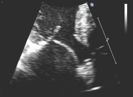 [Figure caption and citation for the preceding image starts]: 肥厚型心肌病伴有收缩期前向运动的心尖三腔心切面由 Samir Kapadia 及 Mehdi H. Shishehbor 提供 [Citation ends].
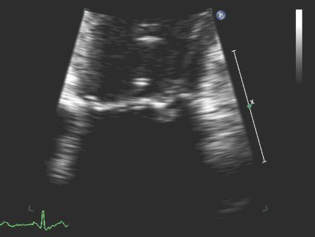 [Figure caption and citation for the preceding image starts]: 连枷二尖瓣后叶的心尖四腔心切面由 Samir Kapadia 及 Mehdi H. Shishehbor 提供 [Citation ends].
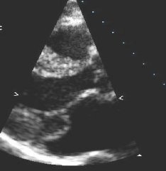 [Figure caption and citation for the preceding image starts]: 胸骨旁长轴图显示二尖瓣后叶脱垂由 Samir Kapadia 及 Mehdi H. Shishehbor 提供 [Citation ends].另外还可用于评估左心室大小和功能的动态变化,以及在症状变化后评价患者。某些较新系统除标准的二维图像外还可生成实时三维图像。
 [Figure caption and citation for the preceding image starts]: 二尖瓣 P2 瓣叶脱垂:二维图由 Prakash P. Punjabi 提供 [Citation ends].
 [Figure caption and citation for the preceding image starts]: 二尖瓣 P2 瓣叶脱垂:三维图由 Prakash P. Punjabi 提供 [Citation ends].
 [Figure caption and citation for the preceding image starts]: 人工二尖瓣伴间歇性二尖瓣反流由 Prakash P. Punjabi 提供 [Citation ends].
 [Figure caption and citation for the preceding image starts]: 人工二尖瓣伴间歇性二尖瓣反流由 Prakash P. Punjabi 提供 [Citation ends].
血流测量
可采用空间映射、血流会聚、肺静脉血流速度模式、过反流口宽度、连续波多普勒密度和形状以及顺行心脏瓣膜血流容量定量进一步评估严重程度。通常使用多种方法的加权平均值。
血流会聚法(也称为近端等速表面积法)包括检查二尖瓣反流图像左心室侧的彩色图像。[10]Enriquez-Sarano M, Avierinos JF, Messika-Zeitoun D, et al. Quantitative determinants of the outcome of asymptomatic mitral regurgitation. N Engl J Med. 2005;352:875-883.http://www.nejm.org/doi/full/10.1056/NEJMoa041451#t=articlehttp://www.ncbi.nlm.nih.gov/pubmed/15745978?tool=bestpractice.com[11]Cosgrove DM, Stewart WJ. Mitral valvuloplasty. Curr Probl Cardiol. 1989;14:359-415.http://www.ncbi.nlm.nih.gov/pubmed/2667897?tool=bestpractice.com[12]Stewart WJ, Currie PJ, Salcedo EE, et al. Evaluation of mitral leaflet motion by echocardiography and jet direction by Doppler color flow mapping to determine the mechanisms of mitral regurgitation. J Am Coll Cardiol. 1992;20:1353-1361.http://www.ncbi.nlm.nih.gov/pubmed/1430686?tool=bestpractice.com[13]Zoghbi WA, Enriquez-Sarano M, Foster E, et al. Recommendations for evaluation of the severity of native valvular regurgitation with two-dimensional and Doppler echocardiography. J Am Soc Echocardiogr. 2003;16:777-802.http://www.ncbi.nlm.nih.gov/pubmed/12835667?tool=bestpractice.com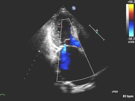 [Figure caption and citation for the preceding image starts]: 1-2+ 向后二尖瓣反流的心尖四腔心切面由 Samir Kapadia 及 Mehdi H. Shishehbor 提供 [Citation ends].
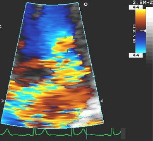 [Figure caption and citation for the preceding image starts]: 4+(重度)二尖瓣反流和大近端等速表面积的心尖四腔心切面由 Samir Kapadia 及 Mehdi H. Shishehbor 提供 [Citation ends].
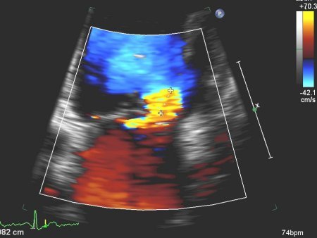 [Figure caption and citation for the preceding image starts]: 严重 MR 伴近端等速表面积的心尖四腔心切面由 Samir Kapadia 及 Mehdi H. Shishehbor 提供 [Citation ends].此血流会聚区是血液加速通过的反流口,这与其从左心室压降到左心房压一致。当此会聚区为半球形时,可通过对其进行分析来评估半球的表面积;因此可计算反流病变的实际大小,这是一个瓣完整性基本参数,其负荷相关性可能比其他方法小。反流口面积超过 0.4 cm^2 表示严重反流,而<0.2 cm^2 为轻度反流。
另外肺静脉血流特征也有助于确定严重程度。[14]Pu M, Griffin BP, Vandervoort PM, et al. The value of assessing pulmonary venous flow velocity for predicting severity of mitral regurgitation: a quantitative assessment integrating left ventricular function. J Am Soc Echocardiogr. 1999;12:736-743.http://www.ncbi.nlm.nih.gov/pubmed/10477418?tool=bestpractice.com正常肺静脉模式的心室收缩期内血流速度高于舒张期内的前向血流速度,这是一个在轻度 MR 病例中持续出现的模式,有时在中度 MR 病例中也出现。由于反流进展,肺静脉收缩期血流速度减慢,收缩期血流速度小于舒张期速度,有时收缩期血流停止。在严重 MR 患者中,收缩期血流往往会逆转,在心室收缩期内血流从左心房流出。这是由于左心房压的大 V 波及在心室收缩期内血流瞬时返回肺实质导致的。
经食管超声心动图
在症状程度与经胸超声心动图检查结果不匹配的罕见情况下可用于更好地评估严重程度及病因。[8]Vahanian A, Alfieri O, Andreotti F, et al; the Joint Task Force on the Management of Valvular Heart Disease of the European Society of Cardiology (ESC) and the European Association for Cardio-Thoracic Surgery (EACTS). Guidelines on the management of valvular heart disease (version 2012). Eur Heart J. 2012;33:2451-2496.http://eurheartj.oxfordjournals.org/content/33/19/2451.full.pdfhttp://www.ncbi.nlm.nih.gov/pubmed/22922415?tool=bestpractice.com[15]Pu M, Vandervoort PM, Griffin BP, et al. Quantification of mitral regurgitation by the proximal convergence method using transesophageal echocardiography. Clinical validation of a geometric correction for proximal flow constraint. Circulation. 1995;92:2169-2177.http://circ.ahajournals.org/content/92/8/2169.fullhttp://www.ncbi.nlm.nih.gov/pubmed/7554198?tool=bestpractice.com应激超声心动图通常可用于确定疾病的严重程度及对患者运动血流动力学的影响。
心电图 (ECG)
所有患者都必须进行心电图 (ECG) 检查,这是一项常规筛查,也可用于检查是否有任何心律失常(例如心房颤动)。[9]Lancellotti P, Moura L, Pierard LA, et al; European Association of Echocardiography. European Association of Echocardiography recommendations for the assessment of valvular regurgitation. Part 2: mitral and tricuspid regurgitation (native valve disease). Eur J Echocardiogr. 2010;11:307-332.http://www.ncbi.nlm.nih.gov/pubmed/20435783?tool=bestpractice.com
导管插管检查及 MRI
伴缺血性心脏病风险的所有患者都应进行心导管插管检查,以检查是否有冠状动脉疾病。[8]Vahanian A, Alfieri O, Andreotti F, et al; the Joint Task Force on the Management of Valvular Heart Disease of the European Society of Cardiology (ESC) and the European Association for Cardio-Thoracic Surgery (EACTS). Guidelines on the management of valvular heart disease (version 2012). Eur Heart J. 2012;33:2451-2496.http://eurheartj.oxfordjournals.org/content/33/19/2451.full.pdfhttp://www.ncbi.nlm.nih.gov/pubmed/22922415?tool=bestpractice.com疑似有肺动脉高压和/或左或右心室功能不良的患者应进行心导管插管检查,以计算肺动脉高压。
左或右心室功能不良和/或二尖瓣环/瓣叶钙化的患者应进行心脏磁共振成像检查。
