临床评价
异物吸入通常在临床上得到诊断,因为大多数患者会出现急性发作的窒息和顽固性咳嗽(窒息危象)。[4]Boyd M, Chatterjee A, Chiles C, et al. Tracheobronchial foreign body aspiration in adults. South Med J. 2009;102:171-174.http://www.ncbi.nlm.nih.gov/pubmed/19139679?tool=bestpractice.com迅速诊断非常重要,尤其对于儿童,因为有研究表明,误吸后两天就医的患者并发症发病率是较早就医患者的两倍。[21]Shlizerman L, Mazzawi S, Rakover Y, et al. Foreign body aspiration in children: the effects of delayed diagnosis. Am J Otolaryngol. 2010;31:320-324.http://www.ncbi.nlm.nih.gov/pubmed/20015771?tool=bestpractice.com
儿童可能表现为进食或玩耍时出现窒息,或药物治疗后咳嗽和哮鸣不见好转。[22]Verghese ST, Hannallah RS. Pediatric otolaryngologic emergencies. Anesthesiol Clin North Am. 2001;2:237-256.http://www.ncbi.nlm.nih.gov/pubmed/11469063?tool=bestpractice.com老年及幼儿患者不能提供提示异物吸入的病史,其症状如咳嗽、喘鸣、呼吸困难等并不具有特异性,常被考虑为其他疾病,如COPD、充血性心力衰竭、哮喘或肺炎等。此时应高度怀疑呼吸道异物的可能性,可作进一步检查以排除或确定诊断。
异物进入气管,可引起剧烈咳嗽(可发出或不能发出声音),和双相喘鸣音(吸气相和呼气相)。气道完全阻塞和窒息可能是由较大物体滞留在气管或喉部中导致的。[22]Verghese ST, Hannallah RS. Pediatric otolaryngologic emergencies. Anesthesiol Clin North Am. 2001;2:237-256.http://www.ncbi.nlm.nih.gov/pubmed/11469063?tool=bestpractice.com呼吸衰竭的早期征象包括呼吸急促、呼吸徐缓、心动过速发展为心动过缓,以及初始增加呼吸功,这可能会进展为呼吸功能下降和不足。发绀,喘鸣和意识水平的改变是不祥的迹象,可预测即将发生呼吸骤停。
有异物吸入病史,有喘鸣、窒息等,体格检查可听到单侧呼吸音减低,局部有喘鸣音,阻塞性肺气肿或肺不张,胸部X线片可见不透明异物影,均提示支气管异物。
胸部 X 射线检查
这是针对疑似异物吸入的病情稳定患者的初始影像学检查方法。应获取标准的正位和侧位视图,以帮助确定物体的位置。[4]Boyd M, Chatterjee A, Chiles C, et al. Tracheobronchial foreign body aspiration in adults. South Med J. 2009;102:171-174.http://www.ncbi.nlm.nih.gov/pubmed/19139679?tool=bestpractice.com若临床上怀疑上累及气道,需加行颈部侧位软组织检查。若患者病情危急,病史和体格检查均高度提示呼吸道异物,则不是必须行胸部X线检查。在这些病例中,应根据需要保护气道,并考虑采用软式或硬式支气管镜检查。
X线上异物的表现不一,因异物的密度、症状持续的时间而异。不透射线物质可在X线上显示,如硬币、图钉、玩具、骨、牙齿及牙科器械等。呼吸道异物中仅2%-19%可见不透射线异物,原因为大多数异物可透射线。此外,X 线照片上所示的不透射线物质可能是黏液嵌塞钙化或支气管结石,因而可能是假阳性结果。[23]Paintal HS, Kuschner WG. Aspiration syndromes: 10 clinical pearls every physician should know. Int J Clin Pract. 2007;61:846-852.http://www.ncbi.nlm.nih.gov/pubmed/17493092?tool=bestpractice.com
诸如肉或蔬菜等有机物质较难显现。[23]Paintal HS, Kuschner WG. Aspiration syndromes: 10 clinical pearls every physician should know. Int J Clin Pract. 2007;61:846-852.http://www.ncbi.nlm.nih.gov/pubmed/17493092?tool=bestpractice.com鳕鱼、黑线鳕和鲑鱼的鱼骨是不透射线的,而鲑鳟鱼、马鲛鱼和鲱鱼的鱼骨则可透射线。[24]Ell SR, Sprigg A. The radio-opacity of fishbones - species variation. Clin Radiol. 1991;44:104-107.http://www.ncbi.nlm.nih.gov/pubmed/1884575?tool=bestpractice.com许多片剂也不可透过X射线。
急诊室针对异物吸入进行的胸部 X 线敏感度仅为 22.6%。[25]Pinto A, Scaglione M, Pinto F. Tracheobronchial aspiration of foreign bodies: current indications for emergency plain chest radiography. Radiol Med. 2006;111:497-506.http://www.ncbi.nlm.nih.gov/pubmed/16779536?tool=bestpractice.com儿童患者检查假阴性率在5%至30%之间,成人患者在8%至80%之间。 其浮动之大可能与吸入异物的物理性质有关。
若异物可透过X射线,胸部X线检查未能发现异物,肺不张、肺炎、空气潴留征、纵隔积气等影像表现可提示气道异物。空气潴留征被认为是早期的影像学表现,由于气道内发生活瓣样阻塞,空气只可进而不可出,空气滞留在肺内表现为空气潴留征。呼气性和吸气性胸部 X 线(若可行)可能有助于检测任何气体滞留。[26]Kavanagh PV, Mason AC, Müller NL. Thoracic foreign bodies in adults. Clin Radiol. 1999;54:353-360.http://www.ncbi.nlm.nih.gov/pubmed/10406334?tool=bestpractice.com对于已吸入异物的儿童,此项结果的阴性预测值为 70%。[10]Righini CA, Morel N, Karkas A, et al. What is the diagnostic value of flexible bronchoscopy in the initial investigation of children with suspected foreign body aspiration? Int J Pediatr Otorhinolaryngol. 2007;71:1383-1390.http://www.ncbi.nlm.nih.gov/pubmed/17580093?tool=bestpractice.com支气管扩张,肺脓肿及脓胸等通常为晚期表现。不同原因引起的中央气道梗阻,如良性或恶性肿瘤,均可引起上述非特异性影像学表现。文献报道约25%的呼吸道异物的病例胸部X线检查未见明显异常,故X线检查正常不能排除异物的可能,还需行进一步的检查。
胸部 CT
对于成人,胸部 CT 可在多达 80% 的病例中检测出胸部 X 线中未显现的异物。[27]Bai W, Zhou X, Gao X, et al. Value of chest CT in the diagnosis and management of tracheobronchial foreign bodies. Pediatr Int. 2011;53:515-518.http://www.ncbi.nlm.nih.gov/pubmed/21129123?tool=bestpractice.com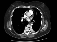 [Figure caption and citation for the preceding image starts]: 胸部CT静脉造影可显示左下肺叶完全塌陷,左侧主支气管内有一不透明异物影,周围有空气影。BMJ病例报道 2008(doi:10.1136/bcr.06.2008.0013).Copyright 2008 BMJ Group Ltd [Citation ends].这种检查方法对具有慢性呼吸系统症状或复发性肺炎的患者尤其有用。可能会发生假阴性 CT 结果(尤其在使用 10 mm 层厚时),在这种情况下可能会遗漏小型物体;此外,对于严重呼吸困难的患者,CT 扫描的图像质量会受到运动伪像的影响。[4]Boyd M, Chatterjee A, Chiles C, et al. Tracheobronchial foreign body aspiration in adults. South Med J. 2009;102:171-174.http://www.ncbi.nlm.nih.gov/pubmed/19139679?tool=bestpractice.comCT表现包括气道管腔内异物的直接征象,以及间接征象如肺不张(62.5%的病例),肺过度透亮(43.75%),支气管扩张(31.25%),肺大叶实变(18.75%),树芽征(18.75%),同侧胸腔积液(18.75%),同侧淋巴结肿大(31.25%),和异物相邻的支气管壁增厚(43.75%)。
胸部 CT 有助于减少对儿科患者进行阴性支气管镜检查的次数。[28]Friedman EM, Anthony B. A five-year analysis of airway foreign body management: toward a better understanding of negative bronchoscopies. Ann Otol Rhinol Laryngol. 2016;125:591-595.http://www.ncbi.nlm.nih.gov/pubmed/26988068?tool=bestpractice.com低剂量多排探测器 CT (multidetector CT, MDCT) 扫描和虚拟支气管镜检查 (virtual bronchoscopy, VB) 的敏感度在 92%-100% 之间,特异度为 80%-85%,但仅限于在有能力进行 VB 的中心使用。[29]Jung SY, Pae SY, Chung SM, et al. Three-dimensional CT with virtual bronchoscopy: a useful modality for bronchial foreign bodies in pediatric patients. Eur Arch Otorhinolaryngol. 2012;269:223-228.http://www.ncbi.nlm.nih.gov/pubmed/21409389?tool=bestpractice.com[30]Bhat KV, Hegde JS, Nagalotimath US, et al. Evaluation of computed tomography virtual bronchoscopy in paediatric tracheobronchial foreign body aspiration. J Laryngol Otol. 2010;124:875-879.http://www.ncbi.nlm.nih.gov/pubmed/20426892?tool=bestpractice.com可能因分泌物和支气管内肿瘤而导致出现假阳性结果。因此,MDCT和VB,如果可用可行,可作为无创性的工具来诊断儿童气道异物,在行支气管镜检查前确定梗阻的确切位置。有研究建议,在出现阳性临床诊断和阴性胸部 X 线时,必须对疑似气管支气管误吸的所有儿童进行 VB,以避免使用硬式支气管镜检查。[30]Bhat KV, Hegde JS, Nagalotimath US, et al. Evaluation of computed tomography virtual bronchoscopy in paediatric tracheobronchial foreign body aspiration. J Laryngol Otol. 2010;124:875-879.http://www.ncbi.nlm.nih.gov/pubmed/20426892?tool=bestpractice.com
对于成人,则应进行支气管镜检查以确认或诊断气道阻塞的其他原因。
支气管镜检查
对于病情稳定的成人,应首先使用软式支气管镜检查以确认异物吸入的疑似病因,并尝试取出异物。[4]Boyd M, Chatterjee A, Chiles C, et al. Tracheobronchial foreign body aspiration in adults. South Med J. 2009;102:171-174.http://www.ncbi.nlm.nih.gov/pubmed/19139679?tool=bestpractice.com颈面部外伤患者,和使用机械通气的患者,均应选择纤维支气管镜为初始检查。在这些患者中,硬质支气管镜更应作为一种治疗方法,而不是作为一种诊断工具。支气管镜检查可确定异物的准确位置,性质,气道阻塞的程度,以及相关的黏膜病变,如黏膜水肿、肉芽组织。尽管大部分病例都建议进行软式支气管镜检查,但对于存在窒息、单侧呼吸音减弱、局部哮鸣、肺部充气过度或肺不张的患者应考虑使用硬式支气管镜检查作为初始诊断和试验治疗。
对于疑似异物吸入的情况稳定儿童,软式支气管镜检查已被证实可安全地确认诊断且可以用于进行治疗。[5]Swanson KL, Prakash UB, Midthun DE, et al. Flexible bronchoscopic management of airway foreign bodies in children. Chest. 2002;121:1695-1700.http://journal.chestnet.org/article/S0012-3692(15)34891-1/fulltexthttp://www.ncbi.nlm.nih.gov/pubmed/12006464?tool=bestpractice.com软式支气管镜检查已被证实能够为大约 12% 患有持续性哮鸣的儿童提供迅速且明确的异物吸入诊断。[31]Cakir E, Ersu RH, Uyan ZS, et al. Flexible bronchoscopy as a valuable tool in the evaluation of persistent wheezing in children. Int J Pediatr Otorhinolaryngol. 2009;73:1666-1668.http://www.ncbi.nlm.nih.gov/pubmed/19733921?tool=bestpractice.com而硬式支气管镜检查则普遍用于为儿童取出异物,[10]Righini CA, Morel N, Karkas A, et al. What is the diagnostic value of flexible bronchoscopy in the initial investigation of children with suspected foreign body aspiration? Int J Pediatr Otorhinolaryngol. 2007;71:1383-1390.http://www.ncbi.nlm.nih.gov/pubmed/17580093?tool=bestpractice.com[32]Farrell P. Rigid bronchoscopy for foreign body removal: anaesthesia and ventilation. Pediatr Anaesth. 2004;14:84-89.http://www.ncbi.nlm.nih.gov/pubmed/14717878?tool=bestpractice.com但由于首次硬式支气管镜检查阴性率高(11% 至 46%),因此对疑似异物吸入的患儿并不推荐首次使用该检查来确诊。事实上,美国胸科学会(American Thoracic Society)将软式支气管镜检查视为针对不确定气管支气管异物病例的具有成本效益的治疗方法,因此应避免不必要地使用硬式支气管镜检查和全身麻醉。[33]Faro A, Wood RE, Schechter MS, et al. Official American Thoracic Society technical standards: flexible airway endoscopy in children. Am J Respir Crit Care Med. 2015;191:1066-1080.http://www.thoracic.org/statements/resources/pldd/pediatric-bronch-ts.pdfhttp://www.ncbi.nlm.nih.gov/pubmed/25932763?tool=bestpractice.com
以下提出了儿童气道异物诊断和处理原则:但是对于某些患者,硬质支气管镜检查应作为初始诊断和治疗手段,如患者已出现窒息,影像学上可见明显异物,或存在与之相关的单侧呼吸音减低、局部喘鸣音、局部肺过度膨胀或肺不张。在所有其他情况下,应先使用软式支气管镜检查进行诊断确认。[10]Righini CA, Morel N, Karkas A, et al. What is the diagnostic value of flexible bronchoscopy in the initial investigation of children with suspected foreign body aspiration? Int J Pediatr Otorhinolaryngol. 2007;71:1383-1390.http://www.ncbi.nlm.nih.gov/pubmed/17580093?tool=bestpractice.com
