多数AVM都是作为对其他疾病如脑内出血(ICH)或癫痫发作行诊断性病情检查的一部分而被诊断的。技术的进步及脑成像技术可用性增加提高了AVMs的检测率,包括有症状的和偶然发现的AVMs。
临床表现
AVMs最常表现为脑内出血(40%-70%)。[2]Al-Shahi R, Warlow C. A systematic review of the frequency and prognosis of arteriovenous malformations of the brain in adults. Brain. 2001;124:1900-1926.https://academic.oup.com/brain/article/124/10/1900/333474/A-systematic-review-of-the-frequency-and-prognosishttp://www.ncbi.nlm.nih.gov/pubmed/11571210?tool=bestpractice.com[27]Brown RD Jr., Wiebers DO, Forbes GS. Unruptured intracranial aneurysms and arteriovenous malformations: frequency of intracranial hemorrhage and relationship of lesions. J Neurosurg. 1990;73:859-863.http://www.ncbi.nlm.nih.gov/pubmed/2230969?tool=bestpractice.comICH的症状和体征与以下情况相关:1)脑实质损伤引起的局灶性神经功能缺损或癫痫发作,或2)血肿本身导致颅内压升高引起的占位效应症状,如剧烈头痛、恶心、呕吐、意识障碍以及昏迷。AVMs是年轻患者自发性脑内血肿的最常见病因。[32]Toffol GJ, Biller J, Adams HP Jr. Nontraumatic intracerebral hemorrhage in young adults. Arch Neurol. 1987;44:483-485.http://www.ncbi.nlm.nih.gov/pubmed/3579658?tool=bestpractice.com出血后第一年再次出血的风险最高,此后年出血率为2%到4%。[18]Stapf C, Mast H, Sciacca RR, et al. Predictors of hemorrhage in patients with untreated brain arteriovenous malformation. Neurology. 2006;66:1350-1355.http://www.ncbi.nlm.nih.gov/pubmed/16682666?tool=bestpractice.com[33]Ondra SL, Troupp H, George ED, et al. The natural history of symptomatic arteriovenous malformations of the brain: a 24-year follow-up. J Neurosurg. 1990;73:387-391.http://www.ncbi.nlm.nih.gov/pubmed/2384776?tool=bestpractice.com[34]Kim H, Sidney S, McCulloch CE, et al. Racial/ethnic differences in longitudinal risk of intracranial hemorrhage in brain arteriovenous malformation patients. Stroke. 2007;38:2430-2437.http://stroke.ahajournals.org/content/38/9/2430.fullhttp://www.ncbi.nlm.nih.gov/pubmed/17673729?tool=bestpractice.com脑内血肿是常见病变,占所有卒中的10%-30%。[35]Ferro JM. Update on intracerebral haemorrhage. J Neurol. 2006;253:985-999.http://www.ncbi.nlm.nih.gov/pubmed/16680558?tool=bestpractice.com在众多的潜在病因中,AVMs占所有脑内出血中的4%。[35]Ferro JM. Update on intracerebral haemorrhage. J Neurol. 2006;253:985-999.http://www.ncbi.nlm.nih.gov/pubmed/16680558?tool=bestpractice.com
未破裂的AVMs常偶然被发现,或可能因局部占位效应、炎症、或“血管盗血”等血流动力学改变而表现出局灶神经系统症状。最常见的后遗症为癫痫,可见于20%的患者。[2]Al-Shahi R, Warlow C. A systematic review of the frequency and prognosis of arteriovenous malformations of the brain in adults. Brain. 2001;124:1900-1926.https://academic.oup.com/brain/article/124/10/1900/333474/A-systematic-review-of-the-frequency-and-prognosishttp://www.ncbi.nlm.nih.gov/pubmed/11571210?tool=bestpractice.com[23]Itoyama Y, Uemura S, Ushio Y, et al. Natural course of unoperated intracranial arteriovenous malformations: study of 50 cases. J Neurosurg. 1989;71:805-809.http://www.ncbi.nlm.nih.gov/pubmed/2585069?tool=bestpractice.com[36]Heros RC, Korosue K, Diebold PM. Surgical excision of cerebral arteriovenous malformations: late results. Neurosurgery. 1990;26:570-577.http://www.ncbi.nlm.nih.gov/pubmed/2330077?tool=bestpractice.com然而,患者也可出现神经功能缺损、头痛、认知减退及血流动力学的紊乱。在新生儿中血流动力学紊乱可能严重到足以引起充血性心力衰竭。
初始检查
脑断层扫描是一项非常有用的检查方法,可以明确或排除脑内出血。AVMs因水肿、占位效应及强化增强而表现为混杂密度区域,从而在CT上可见。25%-30%的患者可伴有钙化。[37]Osborn AG, ed. Diagnostic neuroradiology. St Louis, MO: Mosby; 1994.[38]Riina HA, Gobin YP. Grading and surgical planning for intracranial arteriovenous malformations. Neurosurg Focus. 2001;11:e3.http://www.ncbi.nlm.nih.gov/pubmed/16466235?tool=bestpractice.com如果有证据显示存在血肿,在患者年龄、既往病史的背景下,血肿的形式可影响鉴别诊断及远期管理。 [Figure caption and citation for the preceding image starts]: 左额后部AVM破裂导致的脑内血肿(轴位未强化的CT扫描)来自 R. J. Edwards 先生的收藏;经许可后使用 [Citation ends].
当CT扫描结果阴性且存在AVM相关症状(如癫痫发作)时应行MRI进一步评估。对于中到大的AVMs,MRI的敏感性达80%-95%。[15]Faughnan ME, Palda VA, Garcia-Tsao G, et al. International guidelines for the diagnosis and management of hereditary hemorrhagic telangiectasia. J Med Genet. 2011;48:73-87.http://www.ncbi.nlm.nih.gov/pubmed/19553198?tool=bestpractice.comAVM的血管在神经胶质区(T2加权相)上如存在高速血流表现为“流控效应”,或者如形成血栓或存在湍流则表现为高信号区。各种出血的信号特点取决于出血时间。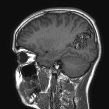 [Figure caption and citation for the preceding image starts]: 左顶枕未破裂的AVM(矢状位T1加权MRI扫描)来自 R. J. Edwards 先生的收藏;经许可后使用 [Citation ends].
 [Figure caption and citation for the preceding image starts]: 左顶枕未破裂的AVM(轴位T2加权MRI扫描)来自 R. J. Edwards 先生的收藏;经许可后使用 [Citation ends].钆强化剂及检测血液成分序列的使用提高了对小AVMs的敏感度。
进一步的检查
数字减影血管造影(DSA)
此为标准的检查方法,可以提供最高的空间分辨率以明确AVM的大小、位置、供血动脉、相关动脉瘤及引流静脉。
其特征性诊断特点为动脉期的静脉充盈(提示动静脉[AV]分流)。但这种现象同样可见于血管肿瘤、外伤、脑实质感染及继发性脑梗。[37]Osborn AG, ed. Diagnostic neuroradiology. St Louis, MO: Mosby; 1994.AVM表现为一块由扩大的供血动脉和扩张、迂曲的引流静脉等异常血管紧密缠绕而成的混杂团块,通常为楔形,指向脑室。 [Figure caption and citation for the preceding image starts]: 脑血管造影(左颈内动脉打药,侧位)显示额叶后部的AVM由胼周动脉供血(小箭头),动脉化的引流静脉(大箭头)向上矢状窦引流。来自 R. J. Edwards 先生的收藏;经许可后使用 [Citation ends].
微动静脉畸形指的是病灶直径<1cm的动静脉畸形,占所有脑动静脉畸形中的7%,其中21%表现为脑内出血。[39]Andreou A, Ioannidis I, Lalloo S, et al. Endovascular treatment of intracranial microarteriovenous malformations. J Neurosurg. 2008;109:1091-1097.http://www.ncbi.nlm.nih.gov/pubmed/19035724?tool=bestpractice.com它们通常是年轻人大的脑内血肿的主要来源,大多数可通过仔细分析血管造影而检测到,其细微的特征性表现为动脉期毛细血管充盈或早期静脉充盈(提示动静脉分流)。然而,有些在血管造影上是隐匿性的。
对于年龄小于60岁,或出血形式不是其他原因(如高血压、淀粉样血管病)的特征性表现,以及能够足够医疗配合去考虑进一步治疗的患者,DSA检查是有必要的。[40]Zhu XL, Chan MS, Poon WS. Spontaneous intracranial hemorrhage: which patients need diagnostic cerebral angiography? A prospective study of 206 cases and review of the literature. Stroke. 1997;28:1406-1409.http://stroke.ahajournals.org/content/28/7/1406.fullhttp://www.ncbi.nlm.nih.gov/pubmed/9227692?tool=bestpractice.com
CT和磁共振血管造影
高清晰的血管成像可通过CT及磁共振获得,同时避免了传统血管造影的风险。该技术在扫描时静脉内予对比剂。获得的图像经过处理,用于显示颅内血管。
它们可以识别大部分动静脉畸形,但对检测小的动静脉分流缺乏敏感性。对于疑似AVM,它们是很有用的辅助检查手段,且可用于治疗后的随访。
四维 CT 血管造影 (CT angiography, CTA) 和磁共振血管造影 (magnetic resonance angiography, MRA) 为传统的 CTA/MRA 增加了时态信息,从而为 AVM 流体动力学提供了一些了解。然而,DSA 仍然是这方面的金标准研究。这些扫描还处于初期阶段,应用并不广泛,但在未来几年可能会得到更多应用。[41]Chandran A, Radon M, Biswas S, et al. Novel use of 4D-CTA in imaging of intranidal aneurysms in an acutely ruptured arteriovenous malformation: is this the way forward? J Neurointerv Surg. 2016;8:e36.http://www.ncbi.nlm.nih.gov/pubmed/26180096?tool=bestpractice.com
功能成像
标准的成像技术如PET、功能性核磁共振或脑磁图可提供功能区(大脑中控制语言、运动功能以及感觉的区域)准确的解剖定位。
功能影像可显示功能上的重要皮质,其范围可能与解剖上的功能区不匹配,这可能是有功能网对于颅内AVMs的适应引起的。[42]Bambakidis NC, Sunshine JL, Faulhaber PF, et al. Functional evaluation of arteriovenous malformations. Neurosurg Focus. 2001;11:e2.http://www.ncbi.nlm.nih.gov/pubmed/16466234?tool=bestpractice.com[43]Lazar RM, Marshall RS, Pile-Spellman J, et al. Anterior translocation of language in patients with left cerebral arteriovenous malformation. Neurology. 1997;49:802-808.http://www.ncbi.nlm.nih.gov/pubmed/9305344?tool=bestpractice.com
超选择性Wada试验
经典的Wada试验将异戊巴比妥钠注入颈内静脉,用于癫痫手术前确立语言和记忆的优势半球。
血管内栓塞术AVMs的供血动脉,常作为手术和放射治疗的辅助手段。在使用硬化剂前,其通过注射异戊巴比妥钠评估潜在的结果而越来越普遍应用。
这项实验显示AVMs可导致脑认知功能的重新分布。[44]Lazar RM. Neuropsychological function and brain arteriovenous malformations: redefining eloquence as a risk for treatment. Neurosurg Focus. 2001;11:e4.http://www.ncbi.nlm.nih.gov/pubmed/16466236?tool=bestpractice.com这是术前评估周围皮质功能最可靠的手段。
正电子发射断层成像(PET)
PET是评估脑组织血流动力学最准确的方法,同时也可提供功能信息。然而,由于资源及所需专业知识所限,其并不易获得使用。[42]Bambakidis NC, Sunshine JL, Faulhaber PF, et al. Functional evaluation of arteriovenous malformations. Neurosurg Focus. 2001;11:e2.http://www.ncbi.nlm.nih.gov/pubmed/16466234?tool=bestpractice.com
实验室检查
脑电图(EEG)
神经心理学
视野检查
