应对所有发生菌血症的患者怀疑可能有感染性心内膜炎,特别是那些有可听得到的心脏杂音的患者。典型的新发或恶化性心脏杂音罕见。闻及新发反流性杂音的患者发生充血性心力衰竭的风险增高。年老者或免疫功能低下患者的表现可能不典型,没有发热。随着人口老龄化,感染性心内膜炎愈发常见,且有临床结局恶化的趋势。[11]Selton-Suty C, Hoen B, Grentzinger A, et al. Clinical and bacteriological characteristics of infective endocarditis in the elderly. Heart. 1997;77:260-263.http://www.ncbi.nlm.nih.gov/pmc/articles/PMC484694/pdf/heart00003-0096.pdfhttp://www.ncbi.nlm.nih.gov/pubmed/9093046?tool=bestpractice.com由于症状表现不明显,对老年人的诊断常常较晚,这无疑使得预后更差。
最初的实验室检查应包括基础的全血细胞计数及电解质检查,间隔1小时的3组血培养和尿液检查。建议开始抗生素治疗前应进行血培养。所有患者都应进行初始心电图检查,随后必须做超声心动图检查。[7]Habib G, Lancellotti P, Antunes MJ, et al. 2015 ESC Guidelines for the management of infective endocarditis. Eur Heart J. 2015;36:3075-3128.http://eurheartj.oxfordjournals.org/content/36/44/3075.longhttp://www.ncbi.nlm.nih.gov/pubmed/26320109?tool=bestpractice.com[20]Habib G, Badano L, Tribouilloy C, et al. Recommendations for the practice of echocardiography in infective endocarditis. Eur J Echocardiogr. 2010;11:202-219.http://www.ncbi.nlm.nih.gov/pubmed/20223755?tool=bestpractice.com [Figure caption and citation for the preceding image starts]: 经胸超声心动图显示三尖瓣前叶和后叶有较大的活动性赘生物Foley JA,Augustine D,Bond R,et al.Lost without Occam's razor:Escherichia coli tricuspid valve endocarditisina non-intravenous drug user.BMJ Case Rep.2010 Aug10;2010.pii:bcr0220102769 [Citation ends].
 [Figure caption and citation for the preceding image starts]: 经食道超声心动图的图像。白色箭头指示患者主动脉瓣膜的赘生物Teoh LS, Hart HH, Soh MC, et al. Bartonella henselae aortic valve endocarditis mimicking systemic vasculitis.BMJ Case Rep. 2010 Oct 21;2010. pii: bcr0420102945 [Citation ends].
急性
患者往往表现出外周或中央栓子的症状和体征,或出现失代偿心衰的证据。因此,任何患者出现头痛伴发热、脑膜刺激征、卒中的症状、胸痛、劳力性呼吸困难、端坐呼吸或夜间阵发性呼吸困难需要立即进行感染性心内膜炎的评估。外周化脓性栓子也可能导致关节痛和背痛。由于发病过程急骤,典型的免疫学特征(例如Osler结、Roth斑)不常见。
亚急性
患者表现为发热和寒战、非特异性全身性症状(盗汗、倦怠、乏力、食欲不振、体重减轻、肌肉痛)或心悸。查体常无特异性发现,但在存在亚急性表现时,更容易得到典型的查体结果,例如 Janeway 皮损(好发于手掌和足底的出血性、斑点性、无痛性斑块),Osler 结节(通常见于指腹或趾腹的小而疼痛的结节性病变),线状出血(通常见于上肢的指甲和下肢的趾甲),或者皮肤坏死。也可有上腭瘀点。眼底镜检查发现罗氏斑(椭圆形、苍白的视网膜病变伴周围出血)。对于有进行性发热和全身性症状的患者,在鉴别诊断时必须总是要考虑到亚急性感染性心内膜炎。 [Figure caption and citation for the preceding image starts]: Janeway损害由英国伦敦大学圣乔治医学院 Sanjay Sharma 提供资料;经许可后使用 [Citation ends].
 [Figure caption and citation for the preceding image starts]: Osler结节由英国伦敦大学圣乔治医学院 Sanjay Sharma 提供资料;经许可后使用 [Citation ends].
 [Figure caption and citation for the preceding image starts]: 皮肤坏死由英国伦敦大学圣乔治医学院 Sanjay Sharma 提供资料;经许可后使用 [Citation ends].
 [Figure caption and citation for the preceding image starts]: Roth斑由英国伦敦大学圣乔治医学院 Sanjay Sharma 提供资料;经许可后使用 [Citation ends].
实验室检查
初始实验室检查应包括基础全血细胞计数及电解质,间隔1小时的3组血培养和尿液检查。检测类风湿因子和评估红细胞沉降率和补体水平也有帮助。
必须在抗生素治疗开始前自不同的静脉穿刺部位获取三份血液培养样本(首份和末份样本应至少间隔 1 小时采集)。[21]Baddour LM, Wilson WR, Bayer AS, et al. Infective endocarditis in adults: diagnosis, antimicrobial therapy, and management of complications: a scientific statement for healthcare professionals from the American Heart Association. Circulation. 2015;132:1435-1486.http://circ.ahajournals.org/content/132/15/1435.fullhttp://www.ncbi.nlm.nih.gov/pubmed/26373316?tool=bestpractice.com培养物不能从留置导管中获取,以使污染风险减至最低。
心电图
因感染的过程可能会导致传导系统病变,故必须进行心电图检查。[22]Porter TR, Airey K, Quader M. Mitral valve endocarditis presenting as complete heart block. Tex Heart Inst J. 2006;33:100-101.http://www.ncbi.nlm.nih.gov/pubmed/16572886?tool=bestpractice.com
超声心动检查
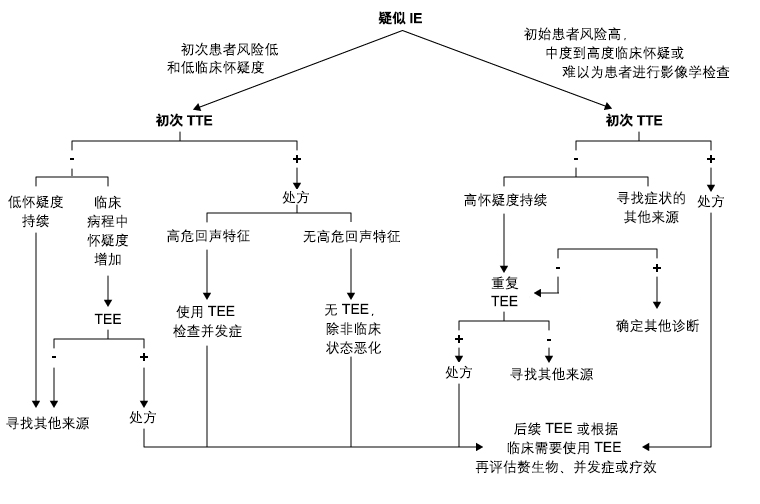
多个指南认为通过经胸超声心动图 (transthoracic echocardiogram, TTE) 往往比经食管超声心动图 (trans-oesophageal echocardiogram, TOE) 进行诊断更为困难。[20]Habib G, Badano L, Tribouilloy C, et al. Recommendations for the practice of echocardiography in infective endocarditis. Eur J Echocardiogr. 2010;11:202-219.http://www.ncbi.nlm.nih.gov/pubmed/20223755?tool=bestpractice.com[21]Baddour LM, Wilson WR, Bayer AS, et al. Infective endocarditis in adults: diagnosis, antimicrobial therapy, and management of complications: a scientific statement for healthcare professionals from the American Heart Association. Circulation. 2015;132:1435-1486.http://circ.ahajournals.org/content/132/15/1435.fullhttp://www.ncbi.nlm.nih.gov/pubmed/26373316?tool=bestpractice.com任何患者,如怀疑有自体瓣膜感染性心内膜炎,应进行经胸超声心动图筛查。如果未发现异常,但仍然怀疑,患者应接受经食道超声心动图检查。经食道超声心动图是怀疑有假体材料的患者有感染性心内膜炎的首选超声心动图检查。在经胸超声心动图结果阳性的患者中,但仍怀疑有并发症或可能有并发症时,且在感染性心内膜炎活动期心脏手术前,经食道超声心动图也适用。 [Figure caption and citation for the preceding image starts]: 超声心动诊断流程。IE (infective endocarditis):感染性心内膜炎;TEE (trans-oesophageal echocardiogram):经食道超声心动图;TTE (transthoracic echocardiogram):经胸超声心动图Baddour, et al. Circulation.2005;经许可后使用 [Citation ends].
计算机断层扫描 (computed tomography, CT)
已经发现计算机断层扫描较经胸超声心动图在检测感染性心内膜炎患者瓣膜异常中存在优势,但它可能发现不了小病灶,例如小的瓣叶穿孔(直径≤2mm)。[23]Feuchtner GM, Stolzmann P, Dichtl W, et al. Multislice computed tomography in infective endocarditis: comparison with transesophageal echocardiography and intraoperative findings. J Am Coll Cardiol. 2009;53:436-444.http://www.ncbi.nlm.nih.gov/pubmed/19179202?tool=bestpractice.com
在符合 Duke 标准定义为“可能的 IE”的患者中,以及证实存在感染性栓塞事件时,SPECT/CT 和 18F-FDG PET/CT 可能尤其有用。[24]Saby L, Laas O, Habib G, et al. Positron emission tomography/computed tomography for diagnosis of prosthetic valve endocarditis: increased valvular 18F-fluorodeoxyglucose uptake as a novel major criterion. J Am Coll Cardiol. 2013;61:2374-2382.http://www.ncbi.nlm.nih.gov/pubmed/23583251?tool=bestpractice.com
磁共振成像 (magnetic resonance imaging, MRI)
磁共振成像 (MRI) 是检查感染性心内膜炎脑部并发症的首选影像学检查方法,多项研究一致报告,在高达 80% 的患者中显示脑梗死。[25]Snygg-Martin U, Gustafsson L, Rosengren L, et al. Cerebrovascular complications in patients with left-sided infective endocarditis are common: a prospective study using magnetic resonance imaging and neurochemical brain damage markers. Clin Infect Dis. 2008;47:23-30.http://cid.oxfordjournals.org/content/47/1/23.longhttp://www.ncbi.nlm.nih.gov/pubmed/18491965?tool=bestpractice.com在未表现出神经系统症状的患者中,MRI 显示 50% 的患者有脑部病变。[26]Hess A, Klein I, Iung B, et al. Brain MRI findings in neurologically asymptomatic patients with infective endocarditis. AJNR Am J Neuroradiol. 2013;34:1579-1584.http://www.ajnr.org/content/34/8/1579.longhttp://www.ncbi.nlm.nih.gov/pubmed/23639563?tool=bestpractice.com重要的是,对于无神经系统症状、但在 MRI 上显示脑部病变的患者,增加了一条额外次要 Duke 标准。因此,最初表现为非确定性 IE 的患者最后可能较早确诊为感染性心内膜炎。[27]Duval X, Iung B, Klein I, et al. Effect of early cerebral magnetic resonance imaging on clinical decisions in infective endocarditis: a prospective study. Ann Intern Med. 2010;152:497-504.http://www.ncbi.nlm.nih.gov/pubmed/20404380?tool=bestpractice.com
重症监护室 (ICU) 中的感染性心内膜炎
ICU 中确诊感染性心内膜炎面临诸多挑战。临床表现多不典型,并可能被其他病变掩盖。此外,因在 ICU 中进行扫描存在实际困难,TTE 诊断的准确性可能更低。因此,必须在诊断过程的更早期阶段考虑 TTE。
 [Figure caption and citation for the preceding image starts]: 经胸超声心动图显示三尖瓣前叶和后叶有较大的活动性赘生物Foley JA,Augustine D,Bond R,et al.Lost without Occam's razor:Escherichia coli tricuspid valve endocarditisina non-intravenous drug user.BMJ Case Rep.2010 Aug10;2010.pii:bcr0220102769 [Citation ends].
 [Figure caption and citation for the preceding image starts]: 经食道超声心动图的图像。白色箭头指示患者主动脉瓣膜的赘生物Teoh LS, Hart HH, Soh MC, et al. Bartonella henselae aortic valve endocarditis mimicking systemic vasculitis.BMJ Case Rep. 2010 Oct 21;2010. pii: bcr0420102945 [Citation ends].
 [Figure caption and citation for the preceding image starts]: Janeway损害由英国伦敦大学圣乔治医学院 Sanjay Sharma 提供资料;经许可后使用 [Citation ends].
 [Figure caption and citation for the preceding image starts]: Osler结节由英国伦敦大学圣乔治医学院 Sanjay Sharma 提供资料;经许可后使用 [Citation ends].
 [Figure caption and citation for the preceding image starts]: 皮肤坏死由英国伦敦大学圣乔治医学院 Sanjay Sharma 提供资料;经许可后使用 [Citation ends].
 [Figure caption and citation for the preceding image starts]: Roth斑由英国伦敦大学圣乔治医学院 Sanjay Sharma 提供资料;经许可后使用 [Citation ends].
 [Figure caption and citation for the preceding image starts]: 超声心动诊断流程。IE (infective endocarditis):感染性心内膜炎;TEE (trans-oesophageal echocardiogram):经食道超声心动图;TTE (transthoracic echocardiogram):经胸超声心动图Baddour, et al. Circulation.2005;经许可后使用 [Citation ends].