早期诊断至关重要,因为越早诊断,长期预后越好。[29]Kinkade S. Testicular cancer. Am Fam Physician. 1999;59:2539-2544,2549-2550.http://www.aafp.org/afp/1999/0501/p2539.htmlhttp://www.ncbi.nlm.nih.gov/pubmed/10323360?tool=bestpractice.com任何睾丸内肿物都要考虑可能是睾丸癌,除非被证实是其他疾病。患者年龄、睾丸癌家族史、睾丸癌既往诊断和最重要的未降睾丸病史,提示可能是睾丸癌。除此之外,根据常规光镜特点和进一步观察或免疫组化进行鉴别诊断有助于得出正确的诊断。[30]Ye H, Ulbright TM. Difficult differential diagnoses in testicular pathology. Arch Pathol Lab Med. 2012;136:435-446.http://www.ncbi.nlm.nih.gov/pubmed/22458906?tool=bestpractice.com
临床评价
典型患者年龄在20-34岁之间。[7]Jemal A, Siegel R, Ward E, et al. Cancer statistics, 2007. CA Cancer J Clin. 2007;57:43-66.http://onlinelibrary.wiley.com/doi/10.3322/canjclin.57.1.43/fullhttp://www.ncbi.nlm.nih.gov/pubmed/17237035?tool=bestpractice.com患者可能主诉没有特异性的睾丸不适,并且在睾丸处可以触摸到一个肿物。症状通常超过2周。约10%的患者主诉突发的疼痛、肿胀(由于相关的血肿或感染)。[31]Dogra V, Bhatt S. Acute painful scrotum. Radiol Clin North Am. 2004;42:349-363.http://www.ncbi.nlm.nih.gov/pubmed/15136021?tool=bestpractice.com
为了早期诊断,对睾丸进行检查是必须的。生殖器查体是一种相对快速和简单的临床评估,当男性患者有生殖器主诉表现时应进行生殖器查体。[32]Marcell AV, Bell DL, Joffe A; Society for Adolescent Health and Medicine, SAHM Male Health Special Interest Group. The male genital examination: a position paper of the Society for Adolescent Health and Medicine. J Adolesc Health. 2012;50:424-425.http://www.ncbi.nlm.nih.gov/pubmed/22443851?tool=bestpractice.com该查体应首先在患者站立位进行,以评估低位睾丸的不对称性。用拇指和其他手指轻柔的触诊睾丸可以确定肿物的位置。肿物应该牢固的附着在睾丸(睾丸内)的一侧或者睾丸下极。应当特别关注肿物的形状和质地。肿物通常是表面光滑的、质硬的和活动度差的。然而,有时也会出现随着肿瘤的轮廓不均匀的变形或均匀的增大。触诊精索是否增厚以及活动度是否良好,需要注意腹股沟是否肿胀。如果有任何疑问,需要行睾丸超声检查。泌尿外科医生应该评估每一个睾丸肿块。
需要进行一个全面的系统检查,因为不论是否有明显的睾丸原发性肿瘤,都有5%-10%的患者会有睾丸外的症状。[33]Richie JP. Neoplasms of the testis. In: Walsh PC, Retik AB, Vaughan ED, et al., eds. Campell's urology. 7th ed. Philadelphia, PA: WB Saunders Company; 2001:2417-2418.这些症状包括骨痛(骨转移)、下肢水肿(静脉栓塞)、锁骨上淋巴结肿大和男性乳房发育症。当腰大肌或神经根侵犯时会出现腰背部疼痛。脊髓和中枢系统转移会导致神经系统症状。
检查
睾丸超声是标准的影像学诊断方法。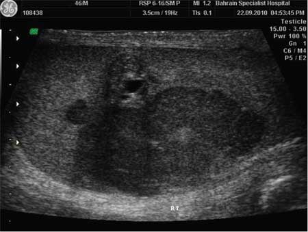 [Figure caption and citation for the preceding image starts]: 睾丸肿物的超声图像表现为低回声团块2011 BMJ病例报道;doi:10.1136/bcr.12.2010.3565 [Citation ends]. 但是,超声检查阴性并不能够排除睾丸癌。彩色多普勒超声可以帮助区分其他良性睾丸肿胀,例如精索静脉曲张。簇状微钙化灶提示睾丸癌的诊断,但是这一特征在许多泌尿外科医生和影像科医生中存在争议。CXR是必须的,用来评估是否存在肺部转移。盆腔和腹部CT扫描用于评估肿瘤播撒的程度。MRI可以作为另一种有用的评估手段。当腹部CT阳性或CXR异常时,建议进行胸部CT扫描。CT-PET扫描并没有被证实优于CT扫描,但是其诊断价值有待进一步研究。
可以应用血清肿瘤标志物来评估分级。[29]Kinkade S. Testicular cancer. Am Fam Physician. 1999;59:2539-2544,2549-2550.http://www.aafp.org/afp/1999/0501/p2539.htmlhttp://www.ncbi.nlm.nih.gov/pubmed/10323360?tool=bestpractice.com升高的β-hCG,甲胎蛋白和LDH的联合评估有助于诊断该病。所有绒毛膜癌患者均会有β-hCG的升高。在胚胎癌、畸胎瘤、卵黄囊肿瘤或混合肿瘤中会出现 AFP 升高。单纯的绒毛膜癌和精原细胞瘤与升高的AFP并不相关。在10%的非精原细胞瘤患者中,LDH是唯一升高的标志物。[34]Sturgeon CM, Duffy MJ, Stenman UH, et al. National Academy of Clinical Biochemistry laboratory medicine practice guidelines for use of tumor markers in testicular, prostate, colorectal, breast, and ovarian cancers. Clin Chem. 2008;54:e11-e79.http://www.clinchem.org/cgi/content/full/54/12/e11http://www.ncbi.nlm.nih.gov/pubmed/19042984?tool=bestpractice.com[35]Stenman UH, Lamerz R, Looijenga LH. Tumor markers in testicular cancers. In: The National Academy of Clinical Biochemistry. Laboratory medicine practice guidelines: use of tumor markers in testicular, prostate, colorectal, breast and ovarian cancers. 2009. http://www.aacc.org/ (last accessed 8 September 2016).https://www.aacc.org/~/media/practice-guidelines/major-tumor-markers/tumormarkersmajor10.pdf?la=en但是,LDH的假阳性率较高,应当主要用于肿瘤负担的评估。胎盘碱性磷酸酶在40%的晚期疾病患者中出现升高。[34]Sturgeon CM, Duffy MJ, Stenman UH, et al. National Academy of Clinical Biochemistry laboratory medicine practice guidelines for use of tumor markers in testicular, prostate, colorectal, breast, and ovarian cancers. Clin Chem. 2008;54:e11-e79.http://www.clinchem.org/cgi/content/full/54/12/e11http://www.ncbi.nlm.nih.gov/pubmed/19042984?tool=bestpractice.com[35]Stenman UH, Lamerz R, Looijenga LH. Tumor markers in testicular cancers. In: The National Academy of Clinical Biochemistry. Laboratory medicine practice guidelines: use of tumor markers in testicular, prostate, colorectal, breast and ovarian cancers. 2009. http://www.aacc.org/ (last accessed 8 September 2016).https://www.aacc.org/~/media/practice-guidelines/major-tumor-markers/tumormarkersmajor10.pdf?la=en三分之一的精原细胞瘤病例 γ-GT 升高。[35]Stenman UH, Lamerz R, Looijenga LH. Tumor markers in testicular cancers. In: The National Academy of Clinical Biochemistry. Laboratory medicine practice guidelines: use of tumor markers in testicular, prostate, colorectal, breast and ovarian cancers. 2009. http://www.aacc.org/ (last accessed 8 September 2016).https://www.aacc.org/~/media/practice-guidelines/major-tumor-markers/tumormarkersmajor10.pdf?la=en50%-70%的非精原细胞型肿瘤患者会出现AFP升高,40%-60%的患者会出现β-hCG的升高。[35]Stenman UH, Lamerz R, Looijenga LH. Tumor markers in testicular cancers. In: The National Academy of Clinical Biochemistry. Laboratory medicine practice guidelines: use of tumor markers in testicular, prostate, colorectal, breast and ovarian cancers. 2009. http://www.aacc.org/ (last accessed 8 September 2016).https://www.aacc.org/~/media/practice-guidelines/major-tumor-markers/tumormarkersmajor10.pdf?la=en[36]Lawton A, Mead G. Staging and prognostic factors in testicular cancer. Semin Surg Oncol. 1999;17:223-229.http://www.ncbi.nlm.nih.gov/pubmed/10588850?tool=bestpractice.com由于肿瘤中合胞体滋养层的出现,精原细胞瘤患者会出现一定的β-hCG升高。
在进行血清肿瘤标志物的检测以后,睾丸癌的诊断建立在对于完整睾丸(睾丸切除术)的切除和检测上。 [Figure caption and citation for the preceding image starts]: 精原细胞瘤由 Francisco G. La Rosa 博士收集,经许可后使用 [Citation ends].
 [Figure caption and citation for the preceding image starts]: 非精原细胞瘤由 Francisco G. La Rosa 博士收集,经许可后使用 [Citation ends].对于该病的所有分期,睾丸切除术是最基础的治疗。对于可疑睾丸肿块的评估,睾丸切除术应该尽早进行。在某些处于早期阶段的患者,睾丸切除术可以治愈该病。
