免疫抑制的程度导致临床表现多样。HIV阳性患者通常能早期诊断,但对于HIV阴性患者,通常会延误卡氏肺孢子虫肺炎(PCP)诊断,这可能出现更严重的后果。支气管肺泡灌洗(BAL)液、诱导痰液或不常使用的肺组织活检中找到病原体可确诊。
病史
当之前有 PCP 病史和/或 CD4 细胞计数<200 个细胞/μl 的 HIV 阳性成人或青少年就诊时,临床医师应当怀疑 PCP,特别是那些进行高效抗逆转录病毒治疗 (HAART) 和 PCP 预防时依从性差的患者。[53]Katz MH, Baron RB, Grady D. Risk stratification of ambulatory patients suspected of Pneumocystis pneumonia. Arch Intern Med. 1991;151:105-110.http://www.ncbi.nlm.nih.gov/pubmed/1985585?tool=bestpractice.com
对于母亲感染HIV而婴儿HIV感染不确定的患者出现呼吸道症状,也应考虑PCP。
一些因以下病史造成免疫功能低下的患者也要考虑PCP:
骨髓移植
实质脏器移植
血液系统肿瘤
长期使用糖皮质激素±免疫抑制剂[24]Bienvenu AL, Traore K, Plekhanova I, et al. Pneumocystis pneumonia suspected cases in 604 non-HIV and HIV patients. Int J Infect Dis. 2016;46:11-17.http://www.ncbi.nlm.nih.gov/pubmed/27021532?tool=bestpractice.com[33]Kovacs JA, Hiemenz JW, Macher AM, et al. Pneumocystis carinii pneumonia: a comparison between patients with the acquired immunodeficiency syndrome and patients with other immunodeficiencies. Ann Intern Med. 1984 May;100(5):663-71.http://www.ncbi.nlm.nih.gov/pubmed/6231873?tool=bestpractice.com
对出现非典型肺炎症状的患者进行HIV感染高危因素评估和HIV检测有助于明确感染PCP的风险。
HIV阳性的PCP患者,病情隐匿进展,在几周内表现出进行性的疲劳,发烧,发冷,盗汗,干咳,呼吸困难。其他提示感染PCP的病史包括复发性细菌性肺炎、消瘦和口腔念珠菌病。
HIV阴性的患者,病情进展更快,症状更严重。[33]Kovacs JA, Hiemenz JW, Macher AM, et al. Pneumocystis carinii pneumonia: a comparison between patients with the acquired immunodeficiency syndrome and patients with other immunodeficiencies. Ann Intern Med. 1984 May;100(5):663-71.http://www.ncbi.nlm.nih.gov/pubmed/6231873?tool=bestpractice.com免疫抑制剂的剂量减少或停用可表现出症状。[21]Pareja JGR, Garland R, Koziel H. Use of adjunctive corticosteroids in severe adult non-HIV Pneumocystis carinii pneumonia. Chest. 1998;113:1215-1224.http://www.ncbi.nlm.nih.gov/pubmed/9596297?tool=bestpractice.com
晚期HIV患者较少出现肺外表现;然而,未进行预防治疗的晚期AIDS或HIV阳性患者可出现系统性感染的临床表现,中枢神经系统受累可损伤认知功能,累及胃肠道可出现腹泻。
体格检查
体格检查无特异性表现,可出现发热、呼吸急促和心动过速。胸部检查常无殊,偶可闻及轻微爆裂音。儿童可表现为发绀。肺外表现少见,但晚期AIDS可出现系统性感染症状。
单侧呼吸音减少可能是气胸表现。胸膜性胸痛可能是气胸的症状,在PCP无气胸患者中少见。
检查
临床症状怀疑PCP,且胸部X线符合PCP表现, [Figure caption and citation for the preceding image starts]: 胸部X线显示两肺轻度网状样间质浸润图片由Matthew Gingo(UPMC)提供 [Citation ends].
 [Figure caption and citation for the preceding image starts]: 胸部X线表现为两肺严重的间质浸润伴有肺囊肿图片由Matthew Gingo(UPMC)提供 [Citation ends].进一步诊断可在诱导痰中查找耶氏肺孢子虫。[3]Murray JF, Nadel JA, Mason RJ. Murray and Nadels Textbook of respiratory medicine. 4th ed. 2005, Philadelphia, PA: Elsevier Saunders.[54]HIV clinical manual. Singh N, Shafer RW, Swindells S (eds). ESun Technologies;2003.
 [Figure caption and citation for the preceding image starts]: 支气管肺泡灌洗(BAL)液显微照片示肺孢子虫包囊,Grocott-Gomori氏六胺银改良法染色呈黑色(甲基绿复染)图片由Matthew Gingo(UPMC)提供 [Citation ends].诱导痰检测的敏感性取决于标本质量、应用的染色技术和实验室经验,敏感性约50%-90%。[43]Centers for Disease Control and Prevention; National Institutes of Health; HIV Medicine Association of the Infectious Diseases Society of America. Guidelines for prevention and treatment of opportunistic infections in HIV-infected adults and adolescents. May 2016. http://aidsinfo.nih.gov/guidelines (last accessed 5 June 2016).http://aidsinfo.nih.gov/contentfiles/lvguidelines/adult_oi.pdf[55]Fortun J, Navas E, Marti-Belda P, et al. Pneumocystis carinii pneumonia in HIV-infected patients: diagnostic yield of induced sputum and immunofluorescent stain with monoclonal antibodies. Eur Respir J. 1992;5:665-669.http://www.ncbi.nlm.nih.gov/pubmed/1628723?tool=bestpractice.com[56]Kovacs JA, Ng VL, Masur H, et al. Diagnosis of Pneumocystis carinii pneumonia: improved detection in sputum with use of monoclonal antibodies. N Engl J Med. 1988;318:589-593.http://www.ncbi.nlm.nih.gov/pubmed/2449613?tool=bestpractice.com[57]Midgley J, Parsons P, Leigh TR, et al. Increased sensitivity of immunofluorescence for detection of Pneumocystis carinii. Lancet. 1989;2:1523.http://www.ncbi.nlm.nih.gov/pubmed/2574793?tool=bestpractice.com[58]Ng VL, Virani NA, Chaisson RE, et al. Rapid detection of Pneumocystis carinii using a direct fluorescent monoclonal antibody stain. J Clin Microbiol. 1990;28:2228-2233.http://www.ncbi.nlm.nih.gov/pubmed/1699968?tool=bestpractice.com
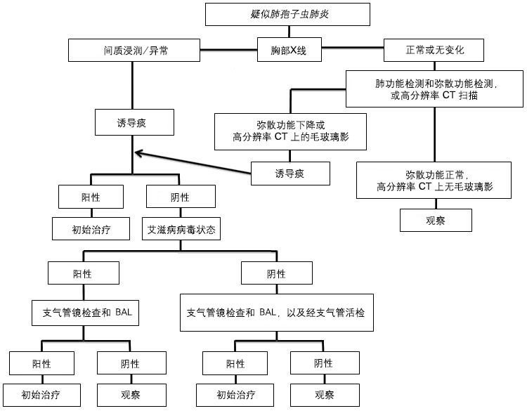
如果诱导痰阴性,可经纤维支气管镜检查支气管肺泡灌洗(BAL)液。HIV阳性患者BAL的敏感率为90%-99%,通常未经纤维支气管镜行活检者可行BAL。[43]Centers for Disease Control and Prevention; National Institutes of Health; HIV Medicine Association of the Infectious Diseases Society of America. Guidelines for prevention and treatment of opportunistic infections in HIV-infected adults and adolescents. May 2016. http://aidsinfo.nih.gov/guidelines (last accessed 5 June 2016).http://aidsinfo.nih.gov/contentfiles/lvguidelines/adult_oi.pdf通常,如果HIV患者BAL阴性,应考虑其他病因,并暂停治疗。
BAL阴性但临床症状高度怀疑PCP或有其他诊断依据,可行纤维支气管镜活检证实。纤维支气管镜活检的敏感率95%-100%。[43]Centers for Disease Control and Prevention; National Institutes of Health; HIV Medicine Association of the Infectious Diseases Society of America. Guidelines for prevention and treatment of opportunistic infections in HIV-infected adults and adolescents. May 2016. http://aidsinfo.nih.gov/guidelines (last accessed 5 June 2016).http://aidsinfo.nih.gov/contentfiles/lvguidelines/adult_oi.pdfHIV阴性的患者由于BAL敏感性低,往往需经纤维支气管镜活检确认诊断。纤维支气管镜活检可出现严重并发症,如出血和气胸。HIV阳性患者行纤维支气管镜活检气胸发生率约9%,其中5%的患者需要插入胸腔引流管。[59]Harcup C, Baier HJ, Pitchenik AE. Evaluation of patients with the acquired immunodeficiency syndrome (AIDS) by fiberoptic bronchoscopy. Endoscopy. 1985;17:217-220.http://www.ncbi.nlm.nih.gov/pubmed/3877629?tool=bestpractice.com
如果临床高度怀疑PCP且之前检查无法确定病因,可行开放性肺活检明确PCP或其他疾病是否存在。
血清LDH的升高(大于220IU/L)具有诊断价值并与预后相关,LDH越高,死亡率越高。[60]Zaman MK, White DA. Serum lactate dehydrogenase levels and Pneumocystis carinii pneumonia. Diagnostic and prognostic significance. Lancet. 1988 Nov 5;2(8619):1049-51.http://www.ncbi.nlm.nih.gov/pubmed/3258483?tool=bestpractice.com可行动脉血气检查是否存在肺泡-动脉(A-a)氧分压差的提高。
患者有PCP的临床表现但无胸片的改变或胸片正常,应该做胸部高分辨率扫描CT,或通过测定一氧化碳扩散功能检测肺功能。如果胸部高分辨率扫描CT显示毛玻璃样变, [Figure caption and citation for the preceding image starts]: 胸部CT扫描显示两肺间质性浸润和肺囊肿,这是典型的卡氏肺孢子虫肺炎(PCP)表现图片由Matthew Gingo(UPMC)提供 [Citation ends].或一氧化碳扩散功能测试是减少的,可进一步行痰诱导检查,如果痰诱导阴性,可行BAL。 尽管CT显示毛玻璃样变或一氧化碳扩散功能测试降低对诊断具有敏感性,但并不是PCP的特异性检查。[61]Gruden JF, Huang L, Turner J, et al. High-resolution CT in the evaluation of clinically suspected Pneumocystis carinii pneumonia in AIDS patients with normal, equivocal, or nonspecific radiographic findings. AJR Am J Roentgenol. 1997;169:967-975.http://www.ajronline.org/doi/pdf/10.2214/ajr.169.4.9308446http://www.ncbi.nlm.nih.gov/pubmed/9308446?tool=bestpractice.com[62]Huang L, Stansell J, Osmond D, et al. Performance of an algorithm to detect Pneumocystis carinii pneumonia in symptomatic HIV-infected persons. Chest. 1999 Apr;115(4):1025-32.http://journal.chestnet.org/article/S0012-3692(16)37737-6/fulltexthttp://www.ncbi.nlm.nih.gov/pubmed/10208204?tool=bestpractice.com[63]Sankary RM, Turner J, Lipavsky A, et al. Alveolar-capillary block in patients with AIDS and Pneumocystis carinii pneumonia. Am Rev Respir Dis. 1988;137:443-449.http://www.ncbi.nlm.nih.gov/pubmed/3257662?tool=bestpractice.com如果诱导痰和BAL阴性,应考虑存在其他病因,并行相应的检查以确诊。如果所有诊断标本耶氏肺孢子虫检查都是阴性,那么患者不符合PCP诊断,应对患者继续观察,并考虑其他病因。根据不同的临床情况进行下一步检查。
PCP诊断不排除二次感染及合并感染。患者在不同的情况下可有相似的临床表现,因此在显微镜下确诊PCP是最可信的方法,临床症状提示PCP时可行经验性治疗,特别是在无法进行诱导痰和BAL检查的地方。
通常通过染色识别痰液或 BAL(支气管肺泡灌洗)液中的金罗维氏肺囊虫,如甲苯胺蓝、吉姆萨染色、D-F 和环六亚甲基四胺银染色。 [Figure caption and citation for the preceding image starts]: 支气管肺泡灌洗(BAL)液显微照片示肺孢子虫包囊,Grocott-Gomori氏六胺银改良法染色呈黑色(甲基绿复染)图片由Matthew Gingo(UPMC)提供 [Citation ends].也可以使用具有更高敏感性的免疫荧光检测(直接荧光抗原检测)。[64]Shelhamer JH, Gill VJ, Quinn TC, et al. The laboratory evaluation of opportunistic pulmonary infections. Ann Intern Med. 1996;124:585-599.http://www.ncbi.nlm.nih.gov/pubmed/8597323?tool=bestpractice.com经支气管活检和BAL可提高诊断率。[65]Broaddus C, Dake MD, Stulbarg MS, et al. Bronchoalveolar lavage and transbronchial biopsy for the diagnosis of pulmonary infections in the acquired immunodeficiency syndrome. Ann Intern Med. 1985;102:747-752.http://www.ncbi.nlm.nih.gov/pubmed/2986505?tool=bestpractice.com
其他检测方法,如PCR,实时荧光PCR,逆转录聚合酶联反应,可以检测低水平的耶氏肺孢子虫DNA。以上检测和S-腺苷蛋氨酸的血浆水平可提高PCP的诊断率,但临床上不常使用。PCR较组织化学染色有较高的假阳性率。[66]Fillaux J, Malvy S, Alvarez M, et al. Accuracy of a routine real-time PCR assay for the diagnosis of Pneumocystis jiroveci pneumonia. J Microbiol Methods. 2008;75:258-261.http://www.ncbi.nlm.nih.gov/pubmed/18606198?tool=bestpractice.com[67]Tuncer S, Erguven S, Kocagoz S, et al. Comparison of cytochemical staining, immunofluorescence and PCR for diagnosis of Pneumocystis carinii on sputum samples. Scand J Infect Dis. 1998;30:125-128.http://www.ncbi.nlm.nih.gov/pubmed/9730296?tool=bestpractice.com[68]Larsen HH, Masur H, Kovacs JA, et al. Development and evaluation of a quantitative, touch-down, real-time PCR assay for diagnosing Pneumocystis carinii pneumonia. J Clin Microbiol. 2002;40:490-494.http://www.ncbi.nlm.nih.gov/pubmed/11825961?tool=bestpractice.com[69]Helweg-Larsen J, Jensen JS, Lundgren B. Diagnostic use of PCR for detection of Pneumocystis carinii in oral wash samples. J Clin Microbiol. 1998;36:2068-2072.http://www.ncbi.nlm.nih.gov/pubmed/9650964?tool=bestpractice.com[70]Larsen HH, Huang L, Kovacs JA, et al. A prospective, blinded study of quantitative touch-down polymerase chain reaction using oral-wash samples for diagnosis of Pneumocystis pneumonia in HIV-infected patients. J Infect Dis. 2004;189:1679-1683.http://www.ncbi.nlm.nih.gov/pubmed/15116305?tool=bestpractice.com[71]Skelly MJ, Holzman RS, Merali S. S-adenosylmethionine levels in the diagnosis of Pneumocystis carinii pneumonia in patients with HIV infection. Clin Infect Dis. 2008;46:467-471.http://cid.oxfordjournals.org/content/46/3/467.longhttp://www.ncbi.nlm.nih.gov/pubmed/18177224?tool=bestpractice.com[72]Summah H, Zhu YG, Falagas ME, et al. Use of real-time polymerase chain reaction for the diagnosis of Pneumocystis pneumonia in immunocompromised patients: a meta-analysis. Chin Med J (Engl). 2013;126:1965-1973.http://www.ncbi.nlm.nih.gov/pubmed/23673119?tool=bestpractice.com
检测血清 (1,3)-β-D-葡聚糖(真菌细胞壁的组成元素)对PCP的诊断也有意义。[73]Watanabe T, Yasuoka A, Tanuma J, et al. Serum (1-->3) beta-D-glucan as a noninvasive adjunct marker for the diagnosis of Pneumocystis pneumonia in patients with AIDS. Clin Infect Dis. 2009;49:1128-1131.http://cid.oxfordjournals.org/content/49/7/1128.longhttp://www.ncbi.nlm.nih.gov/pubmed/19725788?tool=bestpractice.com[74]Shimizu Y, Sunaga N, Dobashi K, et al. Serum markers in interstitial pneumonia with and without Pneumocystis jirovecii colonization: a prospective study. BMC Infect Dis. 2009;9:47.http://www.ncbi.nlm.nih.gov/pmc/articles/PMC2676289/?tool=pubmedhttp://www.ncbi.nlm.nih.gov/pubmed/19383170?tool=bestpractice.com[75]Desmet S, Van Wijngaerden E, Maertens J, et al. Serum (1-3)-beta-D-glucan as a tool for diagnosis of Pneumocystis jirovecii pneumonia in patients with human immunodeficiency virus infection or hematological malignancy. J Clin Microbiol. 2009;47:3871-3874.http://www.ncbi.nlm.nih.gov/pmc/articles/PMC2786638/?tool=pubmedhttp://www.ncbi.nlm.nih.gov/pubmed/19846641?tool=bestpractice.com[76]Del Bono V, Mularoni A, Furfaro E, et al. Clinical evaluation of a (1,3)-beta-D-glucan assay for presumptive diagnosis of Pneumocystis jiroveci pneumonia in immunocompromised patients. Clin Vaccine Immunol. 2009;16:1524-1526.http://www.ncbi.nlm.nih.gov/pmc/articles/PMC2756857/?tool=pubmedhttp://www.ncbi.nlm.nih.gov/pubmed/19692624?tool=bestpractice.com[77]Nakamura H, Tateyama M, Tasato D, et al. Clinical utility of serum beta-D-glucan and KL-6 levels in Pneumocystis jirovecii pneumonia. Intern Med. 2009;48:195-202.https://www.jstage.jst.go.jp/article/internalmedicine/48/4/48_4_195/_articlehttp://www.ncbi.nlm.nih.gov/pubmed/19218768?tool=bestpractice.com有研究显示 PCP 患者血清 (1,3)-β-D-葡聚糖水平明显升高,但在 HIV 阴性 PCP 患者中减少,其水平高低与病情严重性和治疗反应无关联。[78]Huang L. Clinical and translational research in pneumocystis and pneumocystis pneumonia. Parasite. 2011;18:3-11.http://www.ncbi.nlm.nih.gov/pubmed/21395200?tool=bestpractice.com一项研究 (1,3)-β-D-葡聚糖对不同 PCP 人群(HIV 感染和无 HIV 感染)诊断价值的 meta 分析显示,(1,3)-β-D-葡聚糖在诊断方面的混合灵敏度和特异性分别为 96% 和 84%。[79]Onishi A, Sugiyama D, Kogata Y, et al. Diagnostic accuracy of serum 1,3-β-D-glucan for pneumocystis jiroveci pneumonia, invasive candidiasis, and invasive aspergillosis: systematic review and meta-analysis. J Clin Microbiol. 2012;50:7-15.http://jcm.asm.org/content/50/1/7.longhttp://www.ncbi.nlm.nih.gov/pubmed/22075593?tool=bestpractice.com最近的一项 meta 分析显示,在 HIV 感染患者中血清 (1,3)-β-D-葡聚糖水平的敏感性 (92%) 和特异性 (78%) 相似;但是,在非 HIV 感染患者中的敏感性和特异性低,分别为 85% 和 73%。一项纳入HIV感染人数最多的研究中(n=282),69%患有PCP,其敏感性和特异性分别为92%和65%。[80]Sax PE, Komarow L, Finkelman MA, et al. Blood (1->3)-beta-D-glucan as a diagnostic test for HIV-related Pneumocystis jirovecii pneumonia. Clin Infect Dis. 2011;53:197-202.http://cid.oxfordjournals.org/content/53/2/197.longhttp://www.ncbi.nlm.nih.gov/pubmed/21690628?tool=bestpractice.com此外,在一项包括159位AIDS患者研究中,每位患者至少有一种呼吸道症状,其中139位患有PCP,β-葡聚糖的敏感性和特异性(≥80 ng/ml)分别为92.8%和75.0%。[81]Wood BR, Komarow L, Zolopa AR, et al. Test performance of blood beta-glucan for Pneumocystis jirovecii pneumonia in patients with AIDS and respiratory symptoms. AIDS. 2013;27:967-972.http://www.ncbi.nlm.nih.gov/pubmed/23698062?tool=bestpractice.com当无法进行其他敏感性更强的试验时,(1,3)-β-D-葡聚糖的敏感性可能有助于排除 PCP,但其特异性不高,因为 (1,3)-β-D-葡聚糖也可在其他真菌感染中检测出来,其并不是肺孢子虫的特异蛋白,在非 HIV 感染者中的可靠性可能更差。
 [Figure caption and citation for the preceding image starts]: 卡氏肺孢子虫肺炎诊断流程;支气管肺泡灌洗(BAL)Matthew Gingo,改编自辛格,HIV临床手册,2003 [Citation ends].
 [Figure caption and citation for the preceding image starts]: 胸部X线显示两肺轻度网状样间质浸润图片由Matthew Gingo(UPMC)提供 [Citation ends].
 [Figure caption and citation for the preceding image starts]: 胸部X线表现为两肺严重的间质浸润伴有肺囊肿图片由Matthew Gingo(UPMC)提供 [Citation ends].进一步诊断可在诱导痰中查找耶氏肺孢子虫。[3][54]
 [Figure caption and citation for the preceding image starts]: 支气管肺泡灌洗(BAL)液显微照片示肺孢子虫包囊,Grocott-Gomori氏六胺银改良法染色呈黑色(甲基绿复染)图片由Matthew Gingo(UPMC)提供 [Citation ends].诱导痰检测的敏感性取决于标本质量、应用的染色技术和实验室经验,敏感性约50%-90%。[43][55][56][57][58]
 [Figure caption and citation for the preceding image starts]: 胸部CT扫描显示两肺间质性浸润和肺囊肿,这是典型的卡氏肺孢子虫肺炎(PCP)表现图片由Matthew Gingo(UPMC)提供 [Citation ends].或一氧化碳扩散功能测试是减少的,可进一步行痰诱导检查,如果痰诱导阴性,可行BAL。 尽管CT显示毛玻璃样变或一氧化碳扩散功能测试降低对诊断具有敏感性,但并不是PCP的特异性检查。[61][62][63]如果诱导痰和BAL阴性,应考虑存在其他病因,并行相应的检查以确诊。如果所有诊断标本耶氏肺孢子虫检查都是阴性,那么患者不符合PCP诊断,应对患者继续观察,并考虑其他病因。根据不同的临床情况进行下一步检查。
 [Figure caption and citation for the preceding image starts]: 支气管肺泡灌洗(BAL)液显微照片示肺孢子虫包囊,Grocott-Gomori氏六胺银改良法染色呈黑色(甲基绿复染)图片由Matthew Gingo(UPMC)提供 [Citation ends].也可以使用具有更高敏感性的免疫荧光检测(直接荧光抗原检测)。[64]经支气管活检和BAL可提高诊断率。[65]
 [Figure caption and citation for the preceding image starts]: 卡氏肺孢子虫肺炎诊断流程;支气管肺泡灌洗(BAL)Matthew Gingo,改编自辛格,HIV临床手册,2003 [Citation ends].