子宫肌瘤通常在评估月经过多或盆腔痛而行盆腔检查时被发现。盆腔超声是确诊的标准检查。
临床病史
子宫肌瘤诊断的主要病史包括月经过多,盆腔痛(疼痛、压迫或痛经),盆腔包块或不孕。[14]Buttram VC, Reiter RC. Uterine leiomyomata: etiology, symptomatology, and management. Fertil Steril. 1981;36:433-445.http://www.ncbi.nlm.nih.gov/pubmed/7026295?tool=bestpractice.com其他症状可能包括腹胀、泌尿系统症状和便秘。某些患者有高血压、不孕或流产病史。可能有肌瘤家族史。
较为少见的症状为巨大肌瘤压迫周围脏器引起的症状,包括:尿频,更为少见的是尿潴留(继发尿道梗阻),直肠症状,比如便秘和腹部绞痛。巨大肌瘤罕见压迫大血管或影响肺通气。[4]Sciarra J, Dilts PV Jr (eds). Gynecology and obstetrics. Philadelphia, PA: JB Lippincott; 1986.
体格检查
子宫肌瘤双合诊骨盆检查的特征为可触及质韧、增大及不规则的子宫。巨大肿瘤腹腔触诊可触及不规则肿块。[3]Droegemueller W. Benign gynecologic conditions. In: Mishell Jr DR, Stenchever MA, Droegemueller W, et al, eds. Comprehensive gynecology, 3rd edition. St. Louis: Mosby-Yearbook Inc.; 1997:467-516.
超声成像
经腹腔和经阴道超声的盆腔成像是诊断子宫肌瘤最常见的,普遍可用的检查手段。超声可完成包括子宫在内的盆腔脏器的成像,是一项无创的门诊检查项目。[26]Critchley HOD, Warner P, Less AJ, et al. Evaluation of abnormal bleeding: comparison of three outpatient procedures within cohorts defined by age and menopausal status. Health Tech Assess. 2004;8:1-46.http://www.ncbi.nlm.nih.gov/pubmed/15361316?tool=bestpractice.com超声检查便宜、无痛、快速且精确。[27]Hatasaka H. The evaluation of abnormal uterine bleeding. Clin Obstet Gynecol. 2005;48:258-273.http://www.ncbi.nlm.nih.gov/pubmed/15805785?tool=bestpractice.com全面的初步超声检查应包括经阴道和经腹腔两种方式,以确保发现阴道探头无法探查到的子宫病变。[26]Critchley HOD, Warner P, Less AJ, et al. Evaluation of abnormal bleeding: comparison of three outpatient procedures within cohorts defined by age and menopausal status. Health Tech Assess. 2004;8:1-46.http://www.ncbi.nlm.nih.gov/pubmed/15361316?tool=bestpractice.com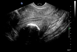 [Figure caption and citation for the preceding image starts]: 单发肌壁间肌瘤经阴道盆腔超声(TVUS)表现为后壁肌层内肿物,位于正常的三层内膜和后壁浆膜之间。来自 M.F.Mitwally 医生和 R.J.Fischer 医生的个人收藏;经许可后使用 [Citation ends].通过经阴道超声更容易发现黏膜下肌瘤,但这些宫腔病变与子宫内膜息肉鉴别仍是一个诊断难题。
 [Figure caption and citation for the preceding image starts]: TVUS显示后壁肌瘤可使宫腔严重变形来自 M.F.Mitwally 医生和 R.J.Fischer 医生的个人收藏;经许可后使用 [Citation ends].
另外,经阴道超声诊断子宫黏膜下肌瘤敏感性100%,特异性94%。特异性稍差是因为经阴道超声难以鉴别子宫内膜息肉与子宫黏膜下肌瘤。[28]Fedele L, Bianchi S, Dorta M, et al. Transvaginal ultrasonography versus hysteroscopy in the diagnosis of uterine submucous myomas. Obstet Gynecol. 1991;77:745-748.http://www.ncbi.nlm.nih.gov/pubmed/2014089?tool=bestpractice.com
子宫内膜活检
子宫内膜癌是发达国家最常见的女性生殖道恶性肿瘤,90%的患者出现有阴道异常出血或异常分泌物。[29]American Congress of Obstetricians and Gynecologists. ACOG practice bulletin number 65: management of endometrial cancer. Obstet Gynecol. 2006;107:952.http://www.ncbi.nlm.nih.gov/pubmed/16582139?tool=bestpractice.com通常,女性患子宫肌瘤发病率高,相当一部分子宫内膜癌患者的阴道异常出血或分泌物与子宫肌瘤相关。因此,不管患者是否有子宫肌瘤,对于异常出血患者,将内膜活检作为最初诊断评估的一部分还是很重要的。
内膜活检诊断子宫内膜癌的精确率约为90%。[30]DiSaia PJ, Creasman WT. Clinical gynecologic oncology, 6th ed. St Louis: Mosby, Inc.; 2002:137-171.5%-10%子宫内膜癌发生于年龄小于40岁女性,因此内膜病变评估对象应包括小于40岁不规则阴道出血和严重阴道出血的患者以及围绝经期(40岁以上)出现异常出血患者。绝经后阴道出血患者应通过内膜组织学检查排除子宫内膜癌。尽管内膜活检阴性,但对于持续不规则阴道出血的患者还应行诊刮。[31]Creasman WT. Malignant tumors of the uterine corpus. In: Rock JA, Jones III HW, eds. Te Linde's operative gynecology, 9th ed. Philadelphia: JB Lippincott Co.; 2003:1445-1486.
盐水灌注宫腔造影超声
宫腔灌注造影超声是检测黏膜下肌壁间肌瘤极为灵敏的辅助检查。[32]Clevenger-Hoeft M, Syrop CH, Stovall DW. Sonohysterography in premenopausal women with and without abnormal bleeding. Obstet Gynecol. 1999;94:516-520.http://www.ncbi.nlm.nih.gov/pubmed/10511351?tool=bestpractice.com
盆腔超声提示宫腔可疑畸形特别是子宫腔本身或宫腔可疑病变时可选择行盐水灌注宫腔造影超声。其还可作为评估绝经前、绝经后女性异常出血和不孕及习惯性流产患者的一线辅助成像检查。[32]Clevenger-Hoeft M, Syrop CH, Stovall DW. Sonohysterography in premenopausal women with and without abnormal bleeding. Obstet Gynecol. 1999;94:516-520.http://www.ncbi.nlm.nih.gov/pubmed/10511351?tool=bestpractice.com[33]American Congress of Obstetricians and Gynecologists. Technology assessment: saline infusion sonohysterography. Int J Gynaecol Obstet. 2004;84:95-98.http://www.ncbi.nlm.nih.gov/pubmed/14968760?tool=bestpractice.com经阴道超声检测宫腔病变时面临的最大问题是鉴别子宫内膜息肉和黏膜下子宫肌瘤;两者均为宫腔占位性病变。子宫内膜息肉完全位于宫腔内且注射盐水时可自由活动。 [Figure caption and citation for the preceding image starts]: 宫腔造影超声提示宫腔内几个小的占位性病变,可疑息肉来自 M.F.Mitwally 医生和 R.J.Fischer 医生的个人收藏;经许可后使用 [Citation ends].
一项纳入52名因良性病而行子宫切除患者的研究显示:经腹腔宫腔造影超声术的灵敏度、特异性和预测值均为100%。[34]Cicinelli E, Romano F, Anastasio PS, et al. Transabdominal sonohysterography, transvaginal sonography, and hysteroscopy in the evaluation of submucous myomas. Obstet Gynecol. 1995;85:42-47.http://www.ncbi.nlm.nih.gov/pubmed/7800322?tool=bestpractice.com8项研究的综述总结了宫腔造影超声诊断黏膜下肌瘤总敏感性94%(91%-100%),特异性95%(88%-100%),高于单纯经阴道超声检查。[35]Dueholm M, Lundorf E, Olesen F. Imaging techniques for evaluation of the uterine cavity and endometrium in premenopausal patients before minimally invasive surgery. Obstet Gynec Survey. 2002;57:389-403.http://www.ncbi.nlm.nih.gov/pubmed/12140373?tool=bestpractice.com综述作者认为宫腔造影超声诊断黏膜下肌瘤可与宫腔镜媲美。然而,另有研究报道宫腔镜诊断黏膜下肌瘤灵敏度100%,特异性96%。这样研究的对照组为经阴道超声检查而不是宫腔造影超声。而且,随着近几年超声技术进步可能会改变该研究的结论。[28]Fedele L, Bianchi S, Dorta M, et al. Transvaginal ultrasonography versus hysteroscopy in the diagnosis of uterine submucous myomas. Obstet Gynecol. 1991;77:745-748.http://www.ncbi.nlm.nih.gov/pubmed/2014089?tool=bestpractice.com
与单纯经阴道或经腹腔盆腔超声检查相比,宫腔造影超声是一种适度侵入性无菌检查,需要经宫颈管放置中等硬度的无菌(约5号French管)进入宫腔。然后注入无菌盐水,高频阴道内探头至少可从两个平面得到即时的超声成像。[33]American Congress of Obstetricians and Gynecologists. Technology assessment: saline infusion sonohysterography. Int J Gynaecol Obstet. 2004;84:95-98.http://www.ncbi.nlm.nih.gov/pubmed/14968760?tool=bestpractice.com少数患者在操作过程中出现不适。[36]Leone FP, Lanzani C, Ferrazzi E. Use of strict sonohysterographic methods for preoperative assessment of submucous myomas. Fertil Steril. 2003;79:998-1002.http://www.ncbi.nlm.nih.gov/pubmed/12749444?tool=bestpractice.com宫腔造影超声禁忌为妊娠、盆腔感染和不明原因盆腔痛。应于检查前行普通盆腔超声检查。[33]American Congress of Obstetricians and Gynecologists. Technology assessment: saline infusion sonohysterography. Int J Gynaecol Obstet. 2004;84:95-98.http://www.ncbi.nlm.nih.gov/pubmed/14968760?tool=bestpractice.com
宫腔造影超声在评估宫腔和内膜下情况包括内膜息肉和黏膜下肌瘤敏感性高达91%-94%,所以很大程度上取代了子宫输卵管造影。[37]Davis PC, O'Neill MJ, Yoder IC, et al. Sonohysterographic findings of endometrial and subendometrial conditions. Radiographics. 2002;22:803-816.http://www.ncbi.nlm.nih.gov/pubmed/12110711?tool=bestpractice.com[38]Gaucherand P, Piacenza JM, Salle B, et al. Sonohysterography of the uterine cavity: preliminary investigations. J Clin Ultrasound. 1995;23:339-348.http://www.ncbi.nlm.nih.gov/pubmed/7673449?tool=bestpractice.com[39]Alatas C, Aksoy E, Akarsu C, et al. Evaluation of intrauterine abnormalities in infertile patients by sonohysterography. Hum Reprod. 1997;12:487-490.http://www.ncbi.nlm.nih.gov/pubmed/9130747?tool=bestpractice.com除了子宫肌瘤,子宫内膜息肉是导致异常出血的常见原因,并且很难与黏膜下肌瘤鉴别。[32]Clevenger-Hoeft M, Syrop CH, Stovall DW. Sonohysterography in premenopausal women with and without abnormal bleeding. Obstet Gynecol. 1999;94:516-520.http://www.ncbi.nlm.nih.gov/pubmed/10511351?tool=bestpractice.com宫腔造影超声敏感性极高(>90%),但有时特异性较低。[35]Dueholm M, Lundorf E, Olesen F. Imaging techniques for evaluation of the uterine cavity and endometrium in premenopausal patients before minimally invasive surgery. Obstet Gynec Survey. 2002;57:389-403.http://www.ncbi.nlm.nih.gov/pubmed/12140373?tool=bestpractice.com与子宫输卵管造影和宫腔镜相比,宫腔造影超声的优点是可更好地确定黏膜下肌瘤在子宫内范围。
宫腔镜
宫腔镜是一种中度侵入性且会引起不适的检查,为确诊宫腔内病变的标准方法。
宫腔镜是诊断子宫内膜息肉很好的方式(敏感性92%),而诊断黏膜下肌瘤并非理想,敏感性仅82%,特异性87%(宫腔造影超声敏感性94%,特异性95%)。[35]Dueholm M, Lundorf E, Olesen F. Imaging techniques for evaluation of the uterine cavity and endometrium in premenopausal patients before minimally invasive surgery. Obstet Gynec Survey. 2002;57:389-403.http://www.ncbi.nlm.nih.gov/pubmed/12140373?tool=bestpractice.com [Figure caption and citation for the preceding image starts]: 宫腔镜检查提示该患者有2个连续黏膜下肌瘤,表现为持续月经过多来自 M.F.Mitwally 医生和 R.J.Fischer 医生的个人收藏;经许可后使用 [Citation ends].
 [Figure caption and citation for the preceding image starts]: 巨大带蒂黏膜下肌瘤的宫腔镜图像来自 M.F.Mitwally 医生和 R.J.Fischer 医生的个人收藏;经许可后使用 [Citation ends].
 [Figure caption and citation for the preceding image starts]: 多发宫腔息肉患者的宫腔镜检查图像,表现为持续阴道淋漓出血来自 M.F.Mitwally 医生和 R.J.Fischer 医生的个人收藏;经许可后使用 [Citation ends].
诊断子宫内膜息肉,宫腔镜极为敏感(>90%),特异性稍差。[35]Dueholm M, Lundorf E, Olesen F. Imaging techniques for evaluation of the uterine cavity and endometrium in premenopausal patients before minimally invasive surgery. Obstet Gynec Survey. 2002;57:389-403.http://www.ncbi.nlm.nih.gov/pubmed/12140373?tool=bestpractice.com
宫腔镜的优点包括可视且能得到手术标本行病理组织学检查。[27]Hatasaka H. The evaluation of abnormal uterine bleeding. Clin Obstet Gynecol. 2005;48:258-273.http://www.ncbi.nlm.nih.gov/pubmed/15805785?tool=bestpractice.com宫腔镜检查对于持续的子宫异常出血且内膜活检阴性的患者非常重要。
磁共振成像
MRI辅助成像在某些特殊情况下是有帮助的,比如,可能与卵巢肿物相似的带蒂浆膜下肌瘤或怀疑为子宫肉瘤时。[40]Thomassin-Naggara I, Darai E, Nassar-Slaba J, et al. Value of dynamic enhanced magnetic resonance imaging for distinguishing between ovarian fibroma and subserous uterine leiomyoma. J Comput Assist Tomogr. 2007;31:236-242.http://www.ncbi.nlm.nih.gov/pubmed/17414760?tool=bestpractice.com[41]Maizlin ZV, Vos PM, Cooperberg PL. Is it a fibroid? Are you sure? Sonography with MRI assistance. Ultrasound. 2007;23:55-62.http://www.ncbi.nlm.nih.gov/pubmed/17558230?tool=bestpractice.comMRI相对昂贵,但能很好评估浆膜下肌瘤在子宫壁内的范围。这对于术前评估哪些子宫肌瘤患者可行微创治疗具有指导意义。 比如,有症状的黏膜下肌瘤患者可行宫腔镜下肌瘤切除。[35]Dueholm M, Lundorf E, Olesen F. Imaging techniques for evaluation of the uterine cavity and endometrium in premenopausal patients before minimally invasive surgery. Obstet Gynec Survey. 2002;57:389-403.http://www.ncbi.nlm.nih.gov/pubmed/12140373?tool=bestpractice.com诊断不明或合并宫颈狭窄或肥大的患者中,MRI可有助于诊断子宫内膜息肉。[42]Wolfman DJ, Ascher SM. Magnetic resonance imaging of benign uterine pathology. Top Magn Reson Imaging. 2006;17:399-407.http://www.ncbi.nlm.nih.gov/pubmed/17417087?tool=bestpractice.com而且,MRI可很好地弥补超声检查的不足之处。[27]Hatasaka H. The evaluation of abnormal uterine bleeding. Clin Obstet Gynecol. 2005;48:258-273.http://www.ncbi.nlm.nih.gov/pubmed/15805785?tool=bestpractice.com
MRI对评估不明确的附件包块很有帮助。[43]Macura KJ. Role of magnetic resonance imaging in the assessment of the female pelvis. Top Magn Reson Imaging. 2006;17:363-364.
因MRI无离子辐射,孕期可安全使用,且能发现常规超声因妊娠子宫增大无法发现的子宫肌瘤。[44]Curtis M, Hopkins MP, Zarlingo T, et al. Magnetic resonance imaging to avoid laparotomy in pregnancy. Obstet Gynecol. 1993; 82:833-836.http://www.ncbi.nlm.nih.gov/pubmed/8414333?tool=bestpractice.com
虽然平滑肌肉瘤在MRI中表现多样,但对其诊断很有帮助,有助于良恶性肌瘤的鉴别。弥漫性平滑肌瘤病也可通过MRI诊断。[45]Kido AK, Togashi K, Koyama T, et al. Diffusely enlarged uterus: evaluation with MR imaging. Radiographics. 2003;23:1423-1439.http://www.ncbi.nlm.nih.gov/pubmed/14615554?tool=bestpractice.com
