尽管技术进步,但诊断仍以临床表现为主要依据,并通过辅助检查进行支持,这些辅助检查包括心电图、胸部 X 线检查、B 型脑利钠肽 (BNP) 和超声心动检查。[1]Ponikowski P, Voors AA, Anker SD, et al. 2016 ESC Guidelines for the diagnosis and treatment of acute and chronic heart failure: the task force for the diagnosis and treatment of acute and chronic heart failure of the European Society of Cardiology (ESC). Eur J Heart Fail. 2016 Aug;18(8):891-975.http://eurheartj.oxfordjournals.org/content/early/2016/05/19/eurheartj.ehw128http://www.ncbi.nlm.nih.gov/pubmed/27207191?tool=bestpractice.com[23]Heart Failure Society of America. Executive summary: HFSA 2010 Comprehensive Heart Failure Practice Guideline. J Card Fail. 2010 Jun;16(6):e1-194.http://www.ncbi.nlm.nih.gov/pubmed/20610207?tool=bestpractice.com 依据临床评估确定左心室 (LV) 收缩功能不全的敏感性约为 81%,但特异性仅为 47%。通过增加心电图检查,可使诊断的特异性可提升至 69%,通过增加胸部 X 线检查可将其提升至 92%。[25]Gillespie ND, McNeill G, Pringle T, et al. Cross sectional study of contribution of clinical assessment and simple cardiac investigations to diagnosis of left ventricular systolic dysfunction in patients admitted with acute dyspnoea. BMJ. 1997 Mar 29;314(7085):936-40.http://www.bmj.com/cgi/content/full/314/7085/936http://www.ncbi.nlm.nih.gov/pubmed/9099117?tool=bestpractice.com
当无创检查和超声心动图图像不理想或怀疑心力衰竭的不常见原因时,以及为了诊断特定的心肌病,可以使用其他检查(例如,使用心导管检查监测血流动力学、计算机体层成像 [CT] 和心脏磁共振 [CMR] 成像)。[1]Ponikowski P, Voors AA, Anker SD, et al. 2016 ESC Guidelines for the diagnosis and treatment of acute and chronic heart failure: the task force for the diagnosis and treatment of acute and chronic heart failure of the European Society of Cardiology (ESC). Eur J Heart Fail. 2016 Aug;18(8):891-975.http://eurheartj.oxfordjournals.org/content/early/2016/05/19/eurheartj.ehw128http://www.ncbi.nlm.nih.gov/pubmed/27207191?tool=bestpractice.com[9]Ezekowitz JA, O'Meara E, McDonald MA, et al. 2017 comprehensive update of the Canadian Cardiovascular Society guidelines for the management of heart failure. Can J Cardiol. 2017 Nov;33(11):1342-1433.https://www.onlinecjc.ca/article/S0828-282X(17)30973-X/fulltexthttp://www.ncbi.nlm.nih.gov/pubmed/29111106?tool=bestpractice.com
病史和危险因素
为了查找急性充血性心力衰竭 (CHF) 的病因(例如心肌缺血、无法控制性高血压、严重的心脏瓣膜病 [包括狭窄和反流]、心律失常、感染、贫血、甲状腺毒症和肺栓塞)以及促使慢性充血性心力衰竭恶化的诱发因素(例如摄入含过多盐分的不当饮食、未遵医嘱用药和过量摄入酒精或药物),应采集详细的病史。临床评分系统有时对诊断急性心力衰竭有帮助。[9]Ezekowitz JA, O'Meara E, McDonald MA, et al. 2017 comprehensive update of the Canadian Cardiovascular Society guidelines for the management of heart failure. Can J Cardiol. 2017 Nov;33(11):1342-1433.https://www.onlinecjc.ca/article/S0828-282X(17)30973-X/fulltexthttp://www.ncbi.nlm.nih.gov/pubmed/29111106?tool=bestpractice.com
症状
心力衰竭表现为呼吸困难、运动耐力降低、下肢肿胀、疲劳和全身乏力。 有时患者会表现出潜在疾病的主要症状,如胸痛、晕厥、心悸或病毒感染前驱症状。
体征
常见体征包括中枢性紫绀、心动过速、颈静脉压升高(JVP)、心尖搏动点移位、第三心音、捻发音或胸腔积液、肝肿大、腹水和水肿。 是否存在这些症状取决于心力衰竭的持续时间、严重程度及根本病因。 晚期心衰患者会出现多数上述症状,而疾病早期则很少出现这些症状。
急性充血性心力衰竭患者在临床上有 4 种血流动力学分类。[26]Nohria A, Lewis E, Stevenson LW. Medical management of advanced heart failure. JAMA. 2002 Feb 6;287(5):628-40.http://jama.ama-assn.org/cgi/content/full/287/5/628http://www.ncbi.nlm.nih.gov/pubmed/11829703?tool=bestpractice.com 这些分类基于血流动力是否存在充盈压升高(湿或干)以及灌注充分或是严重受限(温暖或湿冷),包括:
肺水肿患者表现为严重呼吸窘迫伴随氧饱和度下降(呼吸室内空气时氧饱和度<90%),肺部查体可闻及湿罗音或哮鸣音。 患者出现血液动力异常,收缩压<90mmHg或平均动脉压降低>30mmHg,脉率>60次/分和/或尿量减少(<0.5ml/kg/h),伴或不伴器官淤血,即被认为是心原性休克。
直接检查
对于所有疑似急性充血性心力衰竭的患者,应及时进行心电图和胸部 X 线检查。心电图检查结果一般与潜在疾病相关,包括出现 Q 波、ST-T 段改变、左心室肥厚 (LVH)、左束支传导阻滞和心房颤动。 [Figure caption and citation for the preceding image starts]: 左心室肥大伴窦性心动过速由SyedW.Yusuf,MBBS,MRCPI,和DanielLenihan,MD提供 [Citation ends].
胸部 X 线结果可能显示心脏增大(心胸比率>50%);但心胸比率 (CTR) 与心力衰竭之间的相关性较差,因为舒张性心功能不全的患者、感染性心内膜炎时出现急性瓣膜反流的患者、急性心肌梗死患者的心脏大小往往是正常的。无心力衰竭时也可见心胸比率增大(例如在心包积液和左心室肥厚时)。
肺野评估可显示肺充血的征象,最初在上部区域,然后在水平裂,最后出现肺水肿和胸腔积液。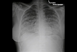 [Figure caption and citation for the preceding image starts]: 急性肺水肿的胸部 X 线征象:肺泡纹理增多,水平裂积液,肋膈角圆钝。由SyedW.Yusuf,MBBS,MRCPI,和DanielLenihan,MD提供 [Citation ends].
 [Figure caption and citation for the preceding image starts]: 急性肺水肿的胸部 X 线显示肺泡纹理增多和双侧胸腔积液由SyedW.Yusuf,MBBS,MRCPI,和DanielLenihan,MD提供 [Citation ends].
X 线检查结果应结合临床情况,因为在某些情况下,肺部浸润和充血有时可能为非心源性原因,例如急性呼吸窘迫综合征 (ARDS) 或肺泡出血。
在少数情况下,胸部 X 线可能显示心包钙化、人工瓣膜或瓣膜钙化。这对识别心力衰竭的潜在可能病因是很有帮助的。
当患者出现疑似急性充血性心力衰竭时,应进行以下血液检查:[23]Heart Failure Society of America. Executive summary: HFSA 2010 Comprehensive Heart Failure Practice Guideline. J Card Fail. 2010 Jun;16(6):e1-194.http://www.ncbi.nlm.nih.gov/pubmed/20610207?tool=bestpractice.com
Hb:判断贫血是否加重心衰。
TFT:甲减和甲亢都会引起心力衰竭。
心肌酶:50% 以上的心源性肺水肿患者(但没有心肌梗死证据)肌钙蛋白 T 水平升高。[27]Perna ER, Macin SM, Parras JI, et al. Cardiac troponin T levels are associated with poor short- and long-term prognosis in patients with acute cardiogenic pulmonary edema. Am Heart J. 2002 May;143(5):814-20.http://www.ncbi.nlm.nih.gov/pubmed/12040342?tool=bestpractice.com 急性充血性心力衰竭患者肌钙蛋白T水平升高可反映由于左心室舒张末压升高而导致的心内膜下心肌缺血。 在急性心源性肺水肿患者中,肌钙蛋白 T 水平≥0.1 μg/L (≥0.1 ng/mL) 是有力的独立预测因素,并与低长期生存率相关。 [27]Perna ER, Macin SM, Parras JI, et al. Cardiac troponin T levels are associated with poor short- and long-term prognosis in patients with acute cardiogenic pulmonary edema. Am Heart J. 2002 May;143(5):814-20.http://www.ncbi.nlm.nih.gov/pubmed/12040342?tool=bestpractice.com一项系统评价和 meta 分析显示,高敏心肌肌钙蛋白 (high sensitive cardiac troponin, hs-cTn) 与偶然心力衰竭的发生密切相关。[28]Evans JDW, Dobbin SJH, Pettit SJ, et al. High-sensitivity cardiac troponin and new-onset heart failure: a systematic review and meta-analysis of 67,063 patients with 4,165 incident heart failure events. JACC Heart Fail. 2018 Mar;6(3):187-97.http://www.ncbi.nlm.nih.gov/pubmed/29331272?tool=bestpractice.com
B型脑钠肽:目前多数医疗中心都会对存在心力衰竭症状的患者进行常规的血清脑钠肽水平检测。 临床评估加入脑钠肽或N-端脑钠肽前体(NT-proBNP)水平的测量,可以显著提高心衰诊断的准确性和治疗的有效性。[29]Maisel AS, Krishnaswamy P, Nowak RM, et al. Rapid measurement of B-type natriuretic peptide in the emergency diagnosis of heart failure. N Engl J Med. 2002 Jul 18;347(3):161-7.http://www.nejm.org/doi/full/10.1056/NEJMoa020233#t=articlehttp://www.ncbi.nlm.nih.gov/pubmed/12124404?tool=bestpractice.com[30]Mueller C, Scholer A, Laule-Kilian K, et al. Use of B-type natriuretic peptide in the evaluation and management of acute dyspnea. N Engl J Med. 2004 Feb 12;350(7):647-54.http://www.nejm.org/doi/full/10.1056/NEJMoa031681#t=articlehttp://www.ncbi.nlm.nih.gov/pubmed/14960741?tool=bestpractice.com B 型脑钠肽对鉴别呼吸困难是心源性还是肺源性也有帮助。[31]Morrison LK, Harrison A, Krishnaswamy P, et al. Utility of a rapid B-natriuretic peptide assay in differentiating congestive heart failure from lung disease in patients presenting with dyspnea. J Am Coll Cardiol. 2002 Jan 16;39(2):202-9.http://www.ncbi.nlm.nih.gov/pubmed/11788208?tool=bestpractice.com 在呼吸困难的患者中,应检测利钠肽生物标志物以协助诊断或排除心力衰竭。[32]Yancy CW, Jessup M, Bozkurt B, et al. 2017 ACC/AHA/HFSA focused update of the 2013 ACCF/AHA guideline for the management of heart failure: a report of the American College of Cardiology/American Heart Association Task Force on Clinical Practice Guidelines and the Heart Failure Society of America. Circulation. 2017 Aug 8;136(6):e137-61.http://circ.ahajournals.org/content/early/2017/04/26/CIR.0000000000000509http://www.ncbi.nlm.nih.gov/pubmed/28455343?tool=bestpractice.com 脑利尿钠肽 (BNP) 水平升高是急性失代偿性心力衰竭患者的住院死亡率的一个预测因子。[33]Fonarow GC, Peacock WF, Phillips CO, et al. Admission B-type natriuretic peptide levels and in-hospital mortality in acute decompensated heart failure. J Am Coll Cardiol. 2007 May 15;49(19):1943-50.http://www.ncbi.nlm.nih.gov/pubmed/17498579?tool=bestpractice.com 然而,BNP 水平升高应结合临床情况考虑,因为它在很多其他疾病中也可能升高,例如心房颤动、肺栓塞或脓毒症。[34]Burke MA, Cotts WG. Interpretation of B-type natriuretic peptide in cardiac disease and other comorbid conditions. Heart Fail Rev. 2007 Mar;12(1):23-36.http://www.ncbi.nlm.nih.gov/pubmed/17345160?tool=bestpractice.com 应在入院时检测利钠肽生物标志物和/或心脏肌钙蛋白的基线水平,以确定急性失代偿性心力衰竭的预后。[32]Yancy CW, Jessup M, Bozkurt B, et al. 2017 ACC/AHA/HFSA focused update of the 2013 ACCF/AHA guideline for the management of heart failure: a report of the American College of Cardiology/American Heart Association Task Force on Clinical Practice Guidelines and the Heart Failure Society of America. Circulation. 2017 Aug 8;136(6):e137-61.http://circ.ahajournals.org/content/early/2017/04/26/CIR.0000000000000509http://www.ncbi.nlm.nih.gov/pubmed/28455343?tool=bestpractice.com
后续的调查
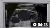
超声心动图是评估急性充血性心力衰竭患者的一个主要手段,应尽快进行。需要该检查来评估心腔大小、心室功能(收缩和舒张)、心室壁厚度、瓣膜功能和心包情况。 [Figure caption and citation for the preceding image starts]: 扩张型心肌病左心室(箭头)的收缩图像;注意相比舒张图像没有变化由SyedW.Yusuf,MBBS,MRCPI,和DanielLenihan,MD提供 [Citation ends].
 [Figure caption and citation for the preceding image starts]: 扩张型心肌病左心室的舒张图像由SyedW.Yusuf,MBBS,MRCPI,和DanielLenihan,MD提供 [Citation ends].
当认为严重冠状动脉病变是心力衰竭的促发因素,或者无法通过其他检查确定急性充血性心力衰竭的原因时,需要进行心导管检查。
心脏 CT 冠状动脉造影是一种无创性检查技术,对于冠状动脉病变验前概率为低中等的心力衰竭患者或者无创性应激试验结果不明确的患者,可用于显示冠状动脉解剖结构,以排除冠状动脉疾病的诊断。
肺动脉 (PA) 导管的常规侵入性血流动力学评估不适用于急性心力衰竭的诊断。 但对于被评估为需要机械循环支持或心脏移植的重度心力衰竭患者,建议采用 PA 导管实施右心插管术,以针对尽管在标准治疗后仍有症状的心力衰竭患者以及血流动力学状态不明的患者确定肺动脉高血压及其可逆性。[1]Ponikowski P, Voors AA, Anker SD, et al. 2016 ESC Guidelines for the diagnosis and treatment of acute and chronic heart failure: the task force for the diagnosis and treatment of acute and chronic heart failure of the European Society of Cardiology (ESC). Eur J Heart Fail. 2016 Aug;18(8):891-975.http://eurheartj.oxfordjournals.org/content/early/2016/05/19/eurheartj.ehw128http://www.ncbi.nlm.nih.gov/pubmed/27207191?tool=bestpractice.com
不建议将心内膜心肌活检用于急性充血性心力衰竭的常规评估,但可用于临床症状提示为急性心肌炎的患者。
 关于如何进行心电图的动画演示
 中心静脉置管的动画演示
当无创检查和超声心动图图像不理想或怀疑心力衰竭的不常见原因时,以及为了诊断特定心肌病,可以使用心脏磁共振成像。[1]Ponikowski P, Voors AA, Anker SD, et al. 2016 ESC Guidelines for the diagnosis and treatment of acute and chronic heart failure: the task force for the diagnosis and treatment of acute and chronic heart failure of the European Society of Cardiology (ESC). Eur J Heart Fail. 2016 Aug;18(8):891-975.http://eurheartj.oxfordjournals.org/content/early/2016/05/19/eurheartj.ehw128http://www.ncbi.nlm.nih.gov/pubmed/27207191?tool=bestpractice.com[9]Ezekowitz JA, O'Meara E, McDonald MA, et al. 2017 comprehensive update of the Canadian Cardiovascular Society guidelines for the management of heart failure. Can J Cardiol. 2017 Nov;33(11):1342-1433.https://www.onlinecjc.ca/article/S0828-282X(17)30973-X/fulltexthttp://www.ncbi.nlm.nih.gov/pubmed/29111106?tool=bestpractice.com
