尽管早期黑素瘤通常无症状,且不易被患者察觉,但应尽量探寻某个可疑色素性损害相关的任何病史。
病史
询问病史的相关问题应该集中在首次观察到皮损的时间上。与直径大于 20 cm 的先天性黑素细胞痣相比,直径小于 20 cm 的先天性黑素细胞痣不太可能发展为黑素瘤。[39]Watt AJ, Kotsis SV, Chung KC. Risk of melanoma arising in large congenital melanocytic nevi: a systematic review. Plast Reconstr Surg. 2004;113:1968-1974.http://www.ncbi.nlm.nih.gov/pubmed/15253185?tool=bestpractice.com尽管在原先存在的痣上发展的病灶不一定都值得怀疑,但是,10% 至 50% 的黑素瘤源自原先存在的黑素细胞痣。[40]Carli P, Massi D, Santucci M, et al. Cutaneous melanoma histologically associated with a nevus and melanoma de novo have a different profile of risk: results from a case-control study. J Am Acad Dermatol. 1999;40:549-557.http://www.ncbi.nlm.nih.gov/pubmed/10188672?tool=bestpractice.com另外,需要询问患者皮损大小,形状和颜色的改变、或者有无溃疡。如果皮损瘙痒或出血,应高度怀疑。[41]Abbasi NR, Shaw HM, Rigel DS, et al. Early diagnosis of cutaneous melanoma: revisiting the ABCD criteria. JAMA. 2004;292:2771-2776.http://www.ncbi.nlm.nih.gov/pubmed/15585738?tool=bestpractice.com无色素的病变称为结节状、无色素、或结缔组织增生性黑素瘤。
某些标志有助于识别高风险的患者:[16]Gandini S, Sera F, Cattaruzza MS, et al. Meta-analysis of risk factors for cutaneous melanoma: III. Family history, actinic damage and phenotypic factors. Eur J Cancer. 2005:41:2040-2059.http://www.ncbi.nlm.nih.gov/pubmed/16125929?tool=bestpractice.com[31]Greene MH, Clark WH Jr, Tucker MA, et al. High risk of malignant melanoma in melanoma-prone families with dysplastic nevi. Ann Intern Med. 1985;102:458-465.http://www.ncbi.nlm.nih.gov/pubmed/3977193?tool=bestpractice.com[34]Elwood JM, Jospson J. Melanoma and sun exposure: an overview of published studies. Int J Cancer. 1997;73:198-203.http://www.ncbi.nlm.nih.gov/pubmed/9335442?tool=bestpractice.com[36]Kanzler MH, Mraz-Gernhard S. Primary cutaneous malignant melanoma and its precursor lesions: diagnostic and therapeutic overview. J Am Acad Dermatol. 2001;45:260-276.http://www.ncbi.nlm.nih.gov/pubmed/11464189?tool=bestpractice.com[42]Psaty EL, Scope A, Halpern AC, et al. Defining the patient at high risk for melanoma. Int J Dermatol. 2010;49:362-376.http://onlinelibrary.wiley.com/doi/10.1111/j.1365-4632.2010.04381.x/fullhttp://www.ncbi.nlm.nih.gov/pubmed/20465687?tool=bestpractice.com
个人或家族黑素瘤病史
个人皮肤癌病史(包括光化损伤)
Fitzpatrick皮肤类型I或II型(浅肤色)
浅色眼睛
雀斑密度高
红色或金色头发
曾经使用过日光浴床
儿童时期晒伤史
较多的黑素细胞痣
有不典型黑素细胞痣(以前称为发育不良痣)
存在巨大先天性黑素细胞痣(>20cm)
具有易患皮肤癌倾向的遗传综合征(例如着色性干皮病)
免疫抑制(例如:免疫抑制剂、大量补骨脂素及UVA[PUVA]疗法的治疗史、或者HIV感染)。
全身症状例如咳嗽、体重减轻、乏力、盗汗及头痛可能是黑素瘤患者发生全身性转移的临床表现。
体格检查
患者应接受整个皮肤表面,包括头皮和黏膜完整的身体检查。美国癌症协会的黑素瘤ABCD迹象法则对医生和患者在早期发现黑色素瘤是有用的。[43]McGovern TW, Litaker MS. Clinical predictors of malignant pigmented lesions. A comparison of the Glasgow seven-point checklist and the American Cancer Society's ABCDs of pigmented lesions. J Dermatol Surg Oncol. 1992;18:22-26.http://www.ncbi.nlm.nih.gov/pubmed/1740563?tool=bestpractice.com建议增加演变 (evolving)(将字母 E 增加至 ABCD 规则)以提高使用 ABCD 规则诊断的敏感性和特异性。[41]Abbasi NR, Shaw HM, Rigel DS, et al. Early diagnosis of cutaneous melanoma: revisiting the ABCD criteria. JAMA. 2004;292:2771-2776.http://www.ncbi.nlm.nih.gov/pubmed/15585738?tool=bestpractice.com
皮损的不对称性
边缘不规则
颜色可变性
直径>6mm
演变。
如果符合以下三个特点中的两点:不规则的轮廓、直径>6mm、且颜色不均匀,则报告黑素瘤的特异性和敏感性分别为0.88和0.73。[43]McGovern TW, Litaker MS. Clinical predictors of malignant pigmented lesions. A comparison of the Glasgow seven-point checklist and the American Cancer Society's ABCDs of pigmented lesions. J Dermatol Surg Oncol. 1992;18:22-26.http://www.ncbi.nlm.nih.gov/pubmed/1740563?tool=bestpractice.com除了黑素瘤ABCDE观察法,相对于正常痣的任何外观不典型的黑素细胞病变均应该引起怀疑。这一单一痣改变的现象被称为“丑小鸭现象”。[44]Grob JJ, Bonerandi JJ. The ugly duckling sign: identification of the common characteristics of nevi in an individual as a basis for melanoma screening. Arch Dermatol. 1998;134:103-104.http://www.ncbi.nlm.nih.gov/pubmed/9449921?tool=bestpractice.com
早期的黑素瘤病灶,尤其是涉及日光暴露部位和肢端部位的病灶,可能完全是斑点样的。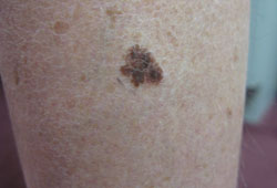 [Figure caption and citation for the preceding image starts]: 浅表扩散性黑素瘤来自于Dr.Hobart Walling和Dr.Brian Swick的个人收藏。 [Citation ends].通常会随着进展出现丘疹样或结节性成分。
 [Figure caption and citation for the preceding image starts]: 结节性黑素瘤来自于Dr.Hobart Walling和Dr.Brian Swick的个人收藏。 [Citation ends].具有非典型特征的黑素瘤,如缺少色素的黑素瘤(无色素性黑素瘤)或类似脂溢性角化病的黑素瘤,如果没有高度怀疑指数很难诊断。结节性黑色素瘤往往会增长迅速,并且没有任何ABCD的特点。在甲床和指甲基质存在色素带的情况下(条纹带黑甲),色素延伸到近端或外侧甲皱襞的现象被称为 Hutchinson 综合征,该综合征与黑素瘤有关。[45]Ronger S, Touzet S, Ligeron C, et al. Dermoscopic examination of nail pigmentation. Arch Dermatol. 2002;138:1327-1333.http://www.ncbi.nlm.nih.gov/pubmed/12374538?tool=bestpractice.com
 [Figure caption and citation for the preceding image starts]: 甲下原位黑素瘤来自于Dr.Hobart Walling和Dr.Brian Swick的个人收藏。 [Citation ends].如果在皮损和引流淋巴结区之间出现了皮下包块,临床上应高度怀疑中途转移的可能。固定的肿大淋巴结与区域淋巴结转移有关。
皮肤血管镜
皮肤血管镜、皮肤镜、皮肤表面显微镜、或表皮荧光显微镜都是利用皮肤血管镜检查皮损的方法。在皮肤镜检查评估中,皮肤病灶被浸液(矿物油或其他液体基质,用于消除表面反射并使表皮和浅表真皮的结构可视化)覆盖。然后手持已连接到光源的放大镜检查皮损。具有偏振光源的皮肤显微镜不需要浸液覆盖。
皮肤镜最重要的应用是辨别良、恶性黑素细胞病变。然而,该技术也可用于从无黑色素皮损中分辨出黑素细胞病变,如脂溢性角化病、色素性基底细胞癌、以及血管病变。
皮肤镜检查中评估的标准包括色素性病变内是否存在色素网、点/球状体、条纹状结构、蓝白盖膜、 [Figure caption and citation for the preceding image starts]: 黑素瘤的“蓝-白色面纱”来自于Dr.Hobart Walling和Dr.Brian Swick的个人收藏。 [Citation ends].斑点、粉刺开口、叶子状色素沉着、红蓝陷窝和色素性病变内的血管结构模式,以及如果存在,边界是否规则。[46]Argenziano G, Soyer HP. Dermoscopy of pigmented skin lesions - a valuable tool for early diagnosis of melanoma. Lancet Oncol. 2001;2:443-449.http://www.ncbi.nlm.nih.gov/pubmed/11905739?tool=bestpractice.com
有各种利用上述皮肤镜检查特征的诊断方法,包括模式分析、[46]Argenziano G, Soyer HP. Dermoscopy of pigmented skin lesions - a valuable tool for early diagnosis of melanoma. Lancet Oncol. 2001;2:443-449.http://www.ncbi.nlm.nih.gov/pubmed/11905739?tool=bestpractice.comABCDE(不对称、边界不规则、颜色变异性、直径>6 mm、演变)规则、[41]Abbasi NR, Shaw HM, Rigel DS, et al. Early diagnosis of cutaneous melanoma: revisiting the ABCD criteria. JAMA. 2004;292:2771-2776.http://www.ncbi.nlm.nih.gov/pubmed/15585738?tool=bestpractice.com[47]Nachbar F, Stolz W, Merkle T, et al. The ABCD rule of dermatoscopy. High prospective value in the diagnosis of doubtful melanocytic skin lesions. J Am Acad Dermatol. 1994;30:551-559.http://www.ncbi.nlm.nih.gov/pubmed/8157780?tool=bestpractice.comMenzies方法,[48]Menzies SW. Surface microscopy of pigmented skin tumors. Australas J Dermatol. 1997;38(suppl 1):S40-S43.http://www.ncbi.nlm.nih.gov/pubmed/10994471?tool=bestpractice.com和七点检查表。[49]Argenziano G, Fabbrocini G, Carli P, et al. Epiluminescence microscopy for the diagnosis of doubtful melanocytic skin lesions. Comparison of the ABCD rule of dermatoscopy and a new 7-point checklist based on pattern analysis. Arch Dermatol. 1998;134:1563-1570.http://jamanetwork.com/journals/jamadermatology/fullarticle/189703http://www.ncbi.nlm.nih.gov/pubmed/9875194?tool=bestpractice.com
与单独肉眼检查相比,训练有素者使用皮肤镜检查增加了诊断的准确性。[50]Vestergaard ME, Macaskill P, Holt PE, et al. Dermoscopy compared with naked eye examination for the diagnosis of primary melanoma: A meta-analysis of studies performed in a clinical setting. Br J Dermatol. 2008;159:669-676.http://www.ncbi.nlm.nih.gov/pubmed/18616769?tool=bestpractice.com利用皮肤镜,诊断黑色素瘤的相对灵敏度提高了19%(用皮肤镜83.2%与不用69.6%),平均特异度提高了6.2%(用皮肤镜85.8%与不用80.8%)。[51]Menzies SW, Zalaudek I. Why perform dermoscopy? The evidence for its role in the routine management of pigmented skin lesions. Arch Dermatol. 2006;142:1111-1112.http://www.ncbi.nlm.nih.gov/pubmed/16983009?tool=bestpractice.com而无正规训练使用皮肤镜检查反而会降低诊断的准确性。[52]Binder M, Puespoeck-Schwart M, Steiner A, et al. Epiluminescence microscopy of small pigmented skin lesions: short-term formal training improves the diagnostic performance of dermatologists. J Am Acad Dermatol. 1997;36:197-202.http://www.ncbi.nlm.nih.gov/pubmed/9039168?tool=bestpractice.com
未来新型的专业诊断手段可能包括在体的共聚焦激光扫描显微镜。[53]Gerger A, Wiltgen M, Langsenlehner U, et al. Diagnostic image analysis of malignant melanoma in in vivo confocal laser-scanning microscopy: a preliminary study. Skin Res Technol. 2008;14:359-363.http://www.ncbi.nlm.nih.gov/pubmed/19159384?tool=bestpractice.com[54]Vestergaard ME, Menzies SW. Automated diagnostic instruments for cutaneous melanoma. Semin Cutan Med Surg. 2008;27:32-36.http://www.ncbi.nlm.nih.gov/pubmed/18486022?tool=bestpractice.com
皮肤活体组织检查
对疑似非典型色素性病变进行活检在诊断中至关重要。任何可疑病灶均应进行活检。对色素性病变行正确的活检可使皮肤病理专家准确诊断黑素瘤,并可影响预后及进一步的手术或其他治疗的范围。理想的活检是对病灶进行完整全层切除。[55]Swanson NA, Lee KK, Gorman A, et al. Biopsy techniques: diagnosis of melanoma. Dermatol Clin. 2002;20:677-680.http://www.ncbi.nlm.nih.gov/pubmed/12380054?tool=bestpractice.com[56]Pardasani AG, Leshin B, Hallman JR, et al. Fusiform incisional biopsy for pigmented skin lesions. Dermatol Surg. 2000;26:622-624.http://www.ncbi.nlm.nih.gov/pubmed/10886267?tool=bestpractice.com[57]Parkinson RW. Shave biopsies - simple and useful. Postgrad Med. 1988;84:161-3,166,169-170.http://www.ncbi.nlm.nih.gov/pubmed/3194325?tool=bestpractice.com
如果整个病变的切除不可行,可以在某些情况下进行全厚度局部活检,包括病灶的任何丘疹或结节状部。然而,此类活检应慎行,因为抽样误差可能导致包括浸润深度在内的组织学标准上的漏诊或误诊。[55]Swanson NA, Lee KK, Gorman A, et al. Biopsy techniques: diagnosis of melanoma. Dermatol Clin. 2002;20:677-680.http://www.ncbi.nlm.nih.gov/pubmed/12380054?tool=bestpractice.com[56]Pardasani AG, Leshin B, Hallman JR, et al. Fusiform incisional biopsy for pigmented skin lesions. Dermatol Surg. 2000;26:622-624.http://www.ncbi.nlm.nih.gov/pubmed/10886267?tool=bestpractice.com
其他检查
血清乳酸脱氢酶
用于根据美国癌症联合会 (AJCC) 黑素瘤分期系统对转移性疾病进行分类。[58]Balch CM, Gershenwald JE, Soong SJ, et al. Final version of 2009 AJCC melanoma staging and classification. J Clin Oncol. 2009;27:6199-6206.http://www.ncbi.nlm.nih.gov/pubmed/19917835?tool=bestpractice.comI期和II黑色素瘤不需检查血清LDH。
胸部X线
1A期患者不需要做胸片检查,但是1B期或更高的需要。而由于胸片是低收益的、假阳性率高、且不易发现早期转移,故胸部X片也是备受争议的。[59]Wang TS, Johnson TM, Cascade PN, et al. Evaluation of staging chest radiographs and serum lactate dehydrogenase for localized melanoma. J Am Acad Dermatol. 2004;51:399-405.http://www.ncbi.nlm.nih.gov/pubmed/15337983?tool=bestpractice.com
CT、MRI或PET
对于I~III期黑素瘤,目前的指南推荐只有存在转移的症状或体征时,才可行CT、MRI或PET检查。然而,在III期、IV期或复发性黑素瘤中,应考虑行胸部、腹部、盆腔CT以及脑部CT或MRI、或是附有脑部成像的全身PET检查。[60]Schwimmer J, Essner R, Patel A, et al. A review of the literature for whole-body FDG PET in the management of patients with melanoma. Q J Nucl Med. 2000;44:153-167.http://www.ncbi.nlm.nih.gov/pubmed/10967625?tool=bestpractice.com[61]Acland KM, O'Doherty MJ, Russell-Jones R. The value of positron emission tomography scanning in the detection of subclinical metastatic melanoma. J Am Acad Dermatol. 2000;42:606-611.http://www.ncbi.nlm.nih.gov/pubmed/10727305?tool=bestpractice.com[62]Ho Shon IA, Chung DK, Saw RP, et al. Imaging in cutaneous melanoma. Nucl Med Commun. 2008;29:847-876.http://www.ncbi.nlm.nih.gov/pubmed/18769303?tool=bestpractice.com
整体而言,PET扫描具有78%~92%的灵敏度及87%~90%特异度,并且对于判断已有区域转移的临床分期是最有效的。[60]Schwimmer J, Essner R, Patel A, et al. A review of the literature for whole-body FDG PET in the management of patients with melanoma. Q J Nucl Med. 2000;44:153-167.http://www.ncbi.nlm.nih.gov/pubmed/10967625?tool=bestpractice.com[61]Acland KM, O'Doherty MJ, Russell-Jones R. The value of positron emission tomography scanning in the detection of subclinical metastatic melanoma. J Am Acad Dermatol. 2000;42:606-611.http://www.ncbi.nlm.nih.gov/pubmed/10727305?tool=bestpractice.com但是,这不足以表明Ⅰ期和Ⅱ期疾病患者的亚临床疾病。[61]Acland KM, O'Doherty MJ, Russell-Jones R. The value of positron emission tomography scanning in the detection of subclinical metastatic melanoma. J Am Acad Dermatol. 2000;42:606-611.http://www.ncbi.nlm.nih.gov/pubmed/10727305?tool=bestpractice.com
在晚期转移性疾病中,CT在肺部成像方面优于PET,而在脑部成像方面MRI优于PET。[62]Ho Shon IA, Chung DK, Saw RP, et al. Imaging in cutaneous melanoma. Nucl Med Commun. 2008;29:847-876.http://www.ncbi.nlm.nih.gov/pubmed/18769303?tool=bestpractice.com
判定中等厚度(Breslow厚度为1.00~3.99mm)黑素瘤患者的临床分期,SLNB(前哨淋巴结活检)优于PET扫描,因为诊断过程中其发生远处转移的风险很低(从局部淋巴结向外移)。[63]El-Maraghi RH, Kielar AZ. PET vs sentinel lymph node biopsy for staging melanoma: a patient intervention, comparison, outcome analysis. J Am Coll Radiol. 2008;5:924-931.http://www.ncbi.nlm.nih.gov/pubmed/18657789?tool=bestpractice.com
前哨淋巴结活检 (Sentinel lymph node biopsy, SLNB)
SLNB 是以肿瘤流向第一个特定淋巴结为基础理念的检查:转移性肿瘤细胞在淋巴结群中遇到的第一个淋巴结。可能有多个引流淋巴结和前哨淋巴结,它取决于个体的淋巴引流模式。
前哨淋巴结活检应在黑素瘤广泛切除时进行。临床上具有局灶黑素瘤的患者行前哨淋巴结活检(SLNB)的适应征包括:Breslow厚度≥1.0mm、或介于0.75mm~1.0mm之间并伴有溃疡;垂直方向上广泛退化≥1.0mm;年轻患者;或高速有丝分裂。[64]Johnson TM, Bradford CR, Gruber SB, et al. Staging workup, sentinel node biopsy, and follow-up tests for melanoma: update or current concepts. Arch Dermatol. 2004;140:107-113.http://www.ncbi.nlm.nih.gov/pubmed/14732667?tool=bestpractice.com[65]Warycha MA, Zakrzewski J, Ni Q, et al. Meta-analysis of sentinel lymph node positivity in thin melanoma (http://www.ncbi.nlm.nih.gov/pubmed/19117354?tool=bestpractice.com
前哨淋巴结的状态是预测生存期最重要且独立的预后因素。[66]Chakera AH, Hesse B, Burak Z, et al. EANM-EORTC general recommendations for sentinel node diagnostics in melanoma. Eur J Nucl Med Mol Imaging. 2009;36:1713-1742.http://www.ncbi.nlm.nih.gov/pubmed/19714329?tool=bestpractice.com[67]Valsecchi ME, Silbermins D, de Rosa N, et al. Lymphatic mapping and sentinel lymph node biopsy in patients with melanoma: a meta-analysis. J Clin Oncol. 2011;29:1479-1487.http://www.ncbi.nlm.nih.gov/pubmed/21383281?tool=bestpractice.com[68]Gershenwald JE, Colome MI, Lee JE, et al. Patterns of recurrence following a negative sentinel lymph node biopsy in 243 patients with stage I or II melanoma. J Clin Oncol. 1998;16:2253-2260.http://www.ncbi.nlm.nih.gov/pubmed/9626228?tool=bestpractice.com[69]Thompson JF, Uren RF. Lymphatic mapping and sentinel node biopsy for melanoma. Expert Rev Anticancer Ther. 2001;1:446-452.http://www.ncbi.nlm.nih.gov/pubmed/12113111?tool=bestpractice.com行SLNB的原因包括确定疾病的分期及预后、早期行淋巴结清扫术、识别相似患者人群参加临床试验、以及考虑是否进行辅助治疗。[70]McMasters KM, Reintgen DS, Ross MI, et al. Sentinel lymph node biopsy for melanoma: controversy despite widespread agreement. J Clin Oncol. 2001;19:2851-2855.http://www.ncbi.nlm.nih.gov/pubmed/11387357?tool=bestpractice.com尽管没有发现明显的总生存优势,多中心选择性淋巴结切除术试验 (MSLT)-Ⅰ的亚组分析显示,SLNB 可提高 1.2 mm 至 3.5 mm 厚度的黑素瘤患者的 10 年远期无病生存率。[71]Morton DL, Thompson JF, Cochran AJ, et al. Final trial report of sentinel-node biopsy versus nodal observation in melanoma. N Engl J Med. 2014;370:599-609.http://www.ncbi.nlm.nih.gov/pubmed/24521106?tool=bestpractice.com
头颈区域的前哨淋巴结活检有技术上的要求,且与身体其他部位相比,识别率较低且假阴性率较高。[72]Tanis PJ, Nieweg OE, van den Brekel MW, et al. Dilemma of clinically node-negative head and neck melanoma: outcome of "watch and wait" policy, elective lymph node dissection, and sentinel node biopsy - a systematic review. Head Neck. 2008;30:380-389.http://www.ncbi.nlm.nih.gov/pubmed/18213724?tool=bestpractice.com[73]de Rosa N, Lyman GH, Silbermins D, et al. Sentinel node biopsy for head and neck melanoma: a systematic review. Otolaryngol Head Neck Surg. 2011;145:375-382.http://www.ncbi.nlm.nih.gov/pubmed/21540313?tool=bestpractice.com
BRAF突变分析
IIIC和IV期肿瘤无法切除的黑素瘤患者可行BRAF基因分析。
大约40%的黑色素瘤含有活化的BRAF突变基因,最常见的位置在V600上。缬氨酸突变为谷氨酸(V600E)占黑素瘤BRAF突变的75%~90%。其他少见的突变包括缬氨酸突变为赖氨酸(V600K)占BRAF突变的10%~30%,和缬氨酸突变为精氨酸(V600R)占1%~3%。这些突变可导致丝分裂原活化蛋白激酶信号通路的活化。[22]Long GV, Menzies AM, Nagrial AM, et al. Prognostic and clinicopathologic associations of oncogenic BRAF in metastatic melanoma. J Clin Oncol. 2011;29:1239-1246.http://ascopubs.org/doi/full/10.1200/jco.2010.32.4327http://www.ncbi.nlm.nih.gov/pubmed/21343559?tool=bestpractice.com[23]Jakob JA, Bassett RL Jr, Ng CS, et al. NRAS mutation status is an independent prognostic factor in metastatic melanoma. Cancer. 2012;118:4014-4023.http://onlinelibrary.wiley.com/doi/10.1002/cncr.26724/fullhttp://www.ncbi.nlm.nih.gov/pubmed/22180178?tool=bestpractice.com[24]Menzies AM, Haydu LE, Visintin L, et al. Distinguishing clinicopathologic features of patients with V600E and V600K BRAF-mutant metastatic melanoma. Clin Cancer Res. 2012;18:3242-3249.http://clincancerres.aacrjournals.org/content/18/12/3242.longhttp://www.ncbi.nlm.nih.gov/pubmed/22535154?tool=bestpractice.com
利用VE1抗体的免疫组织化学通常是检测BRAF方法的首选。对于检测BRAF的V600E,它具有快速、便宜、灵敏度和特异性高的特点。[74]Long GV, Wilmott JS, Capper D, et al. Immunohistochemistry is highly sensitive and specific for the detection of V600E BRAF mutation in melanoma. Am J Surg Pathol. 2013;37:61-65.http://www.ncbi.nlm.nih.gov/pubmed/23026937?tool=bestpractice.com若免疫组织化学结果为阴性,可使用针对稀有突变基因型的分子检测方法。
可用一种自动分子试验方法来检测V600位置上的BRAF突变,但它也只能容易检测V600E突变。[75]Chapman PB, Hauschild A, Robert C, et al. Improved survival with vemurafenib in melanoma with BRAF V600E mutation. N Engl J Med. 2011;364:2507-2516.http://www.nejm.org/doi/full/10.1056/NEJMoa1103782#t=articlehttp://www.ncbi.nlm.nih.gov/pubmed/21639808?tool=bestpractice.com还有其他几个平台可以检测所有已知的V600突变,包括焦磷酸测序技术、新一代测序和Sanger测序。
CDKN2A基因检测
黑素瘤的分期
美国癌症联合委员会(AJCC)黑素瘤分期系统是基于肿瘤、淋巴结、转移(TNM)的分类:[58]Balch CM, Gershenwald JE, Soong SJ, et al. Final version of 2009 AJCC melanoma staging and classification. J Clin Oncol. 2009;27:6199-6206.http://www.ncbi.nlm.nih.gov/pubmed/19917835?tool=bestpractice.com
