若患者存在张力性气胸症状,需立即进行干预。
气胸患者通常自诉存在呼吸困难和胸痛症状。呼吸短促的程度与气胸的大小有关,还(如果存在的话)与预先存在的肺部疾病严重程度有关。通常患者可准确指出症状发作时间。
应评估发生自发性气胸的危险因素,例如慢性呼吸系统疾病。[1]Jantz MA, Pierson DJ. Pneumothorax and barotraumas. Clin Chest Med. 1994;15:75-91.http://www.ncbi.nlm.nih.gov/pubmed/8200194?tool=bestpractice.com 因卡氏肺囊虫肺部感染可导致气胸,故应询问患者是否存在 HIV 感染的危险因素。[23]Wait MA, Estrera A. Changing clinical spectrum of spontaneous pneumothorax. Am J Surg. 1992;164:528-531.http://www.ncbi.nlm.nih.gov/pubmed/1443382?tool=bestpractice.com
临床表现
张力性气胸
患者主诉严重和恶化的呼吸困难,伴快速费力呼吸、紫绀、发汗和心动过速的症状。
需立即对受累半胸进行减压干预。[1]Jantz MA, Pierson DJ. Pneumothorax and barotraumas. Clin Chest Med. 1994;15:75-91.http://www.ncbi.nlm.nih.gov/pubmed/8200194?tool=bestpractice.com[31]Vukich DJ. Diseases of the pleural space. Emerg Med Clin North Am. 1989;7:309-324.http://www.ncbi.nlm.nih.gov/pubmed/2653802?tool=bestpractice.com
 张力性气胸空针减压 (needle decompression) 动画演示
原发性自发性气胸
患者通常主诉呼吸困难突然发作和同侧胸痛症状。呼吸困难的严重程度与进入胸膜间隙的空气量相关。
大部分原发性自发性气胸发生在休息时。因此,患者经常可以回忆起气胸发生的准确时间。[34]O'Neill S. Spontaneous pneumothorax: aetiology, management and complications. Ir Med J. 1987;80:306-311.http://www.ncbi.nlm.nih.gov/pubmed/3325452?tool=bestpractice.com
在某些情况下,胸痛和呼吸困难的典型症状可能比较轻微或者甚至不存在,需保持高度怀疑。
继发性自发性气胸
患者通常主诉呼吸困难和同侧胸痛。因为存在基础肺部疾病,其症状通常比原发性自发性气胸患者更为严重。[31]Vukich DJ. Diseases of the pleural space. Emerg Med Clin North Am. 1989;7:309-324.http://www.ncbi.nlm.nih.gov/pubmed/2653802?tool=bestpractice.com
月经性气胸是继发性自发性气胸。通常发生于右侧。若考虑到这种可能性,诊断并不困难。
阻塞性肺不张后气胸
体格检查
张力性气胸
查体发现与严重自发性气胸患者相似;但受累的半侧胸廓更大,肋间间隙变宽且气管移向对侧胸廓。
患者心肺功能突然恶化通常预示着张力性气胸的发生。因为血液流向大脑受阻,很快会发生意识丧失。[1]Jantz MA, Pierson DJ. Pneumothorax and barotraumas. Clin Chest Med. 1994;15:75-91.http://www.ncbi.nlm.nih.gov/pubmed/8200194?tool=bestpractice.com
原发性自发性气胸
继发性自发性气胸
查体发现与原发性自发性气胸患者相同;然而,因为患者患有基础呼吸疾病,所以查体结果的可靠性较低。[1]Jantz MA, Pierson DJ. Pneumothorax and barotraumas. Clin Chest Med. 1994;15:75-91.http://www.ncbi.nlm.nih.gov/pubmed/8200194?tool=bestpractice.com 检查也可能发现与基础呼吸系统疾病相关的结果。
月经性气胸是继发性自发性气胸。通常发生于右侧。若考虑到这种可能性,诊断并不困难。
阻塞性肺不张后气胸
症状包括呼吸音减弱和叩诊时反响过强。然而,胸廓并无过度扩张。[3]Berdon WE, Dee GJ, Abramson ST, et al. Localized pneumothorax adjacent to a collapsed lobe: a sign of bronchial obstruction. Radiology. 1984;150:691-694.http://www.ncbi.nlm.nih.gov/pubmed/6695068?tool=bestpractice.com
影像学检查
张力性气胸是一种急症。若临床怀疑张力性气胸,应立即对受累胸廓进行减压。不应浪费时间等待放射照相结果的确认。医学干预的推迟可能会导致患者死亡。[1]Jantz MA, Pierson DJ. Pneumothorax and barotraumas. Clin Chest Med. 1994;15:75-91.http://www.ncbi.nlm.nih.gov/pubmed/8200194?tool=bestpractice.com
胸部 X 线一般被推荐作为其他大多数气胸的一线检查方法,可显示脏胸膜线。[35]O'Connor AR, Morgan WE. Radiological review of pneumothorax. BMJ. 2005;330:1493-1497.http://www.ncbi.nlm.nih.gov/pmc/articles/PMC558461/?tool=pubmedhttp://www.ncbi.nlm.nih.gov/pubmed/15976424?tool=bestpractice.com 不建议将呼气 X 光相片用于常规诊断。[35]O'Connor AR, Morgan WE. Radiological review of pneumothorax. BMJ. 2005;330:1493-1497.http://www.ncbi.nlm.nih.gov/pmc/articles/PMC558461/?tool=pubmedhttp://www.ncbi.nlm.nih.gov/pubmed/15976424?tool=bestpractice.com 在继发性自发性气胸患者中可能很难看到胸膜线,因为临近的患肺可能会过度透亮(例如,肺气肿改变患者)。另外,很难鉴别大范围、薄壁大疱和气胸。这种情况下,获得胸部 CT 结果确诊疾病可能十分必要。[35]O'Connor AR, Morgan WE. Radiological review of pneumothorax. BMJ. 2005;330:1493-1497.http://www.ncbi.nlm.nih.gov/pmc/articles/PMC558461/?tool=pubmedhttp://www.ncbi.nlm.nih.gov/pubmed/15976424?tool=bestpractice.com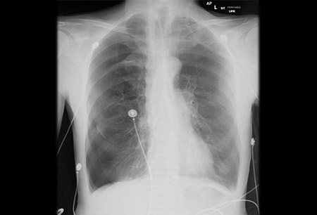 [Figure caption and citation for the preceding image starts]: 前后位胸部 X 线显示右侧气胸由 Ryland P. Byrd 医生提供 [Citation ends].
对于阻塞性肺不张后气胸,除存在脏胸膜线外,还存在同侧体积减小和肺不张症状。CT 可能表明支气管梗阻的存在。
因为在多种临床环境下超声检查更为普遍,这种影像学形式作为临床诊断气胸的方式也越来越受欢迎。[36]Volpicelli G. Sonographic diagnosis of pneumothorax. Intensive Care Med. 2011;37:224-232.http://www.ncbi.nlm.nih.gov/pubmed/21103861?tool=bestpractice.com 对于有经验的医生,超声在诊断气胸方面的敏感度和特异度比较合理。[37]Lichtenstein DA, Meziere GA. Relevance of lung ultrasound in the diagnosis of acute respiratory failure: the BLUE protocol. Chest. 2008;134:117-125.http://www.ncbi.nlm.nih.gov/pubmed/18403664?tool=bestpractice.com[38]Lichtenstein DA, Meziere GA, Lascols N, et al. Ultrasound diagnosis of occult pneumothorax. Crit Care Med. 2005;33:1231-1238.http://www.ncbi.nlm.nih.gov/pubmed/15942336?tool=bestpractice.com 胸部超声检查在发现不能行动的成人钝性胸部创伤患者的气胸方面似乎似乎有用。[39]Wilkerson RG, Stone MB. Sensitivity of bedside ultrasound and supine anteroposterior chest radiographs for the identification of pneumothorax after blunt trauma. Acad Emerg Med. 2010;17:11-17.http://onlinelibrary.wiley.com/doi/10.1111/j.1553-2712.2009.00628.x/fullhttp://www.ncbi.nlm.nih.gov/pubmed/20078434?tool=bestpractice.com 一些研究者建议将超声作为高级创伤生命支持 (ATLS) 方法的辅助手段。[40]Abdulrahman Y, Musthafa S, Hakim SY, et al. Utility of extended FAST in blunt chest trauma: is it the time to be used in the ATLS algorithm? World J Surg. 2015;39:172-178.http://www.ncbi.nlm.nih.gov/pubmed/25205343?tool=bestpractice.com
检测气胸时,胸部 CT 扫描比胸片或超声检查更加灵敏。[41]Sharma A, Jindal P. Principles of diagnosis and management of traumatic pneumothorax. J Emerg Trauma Shock. 2008;1:34-41.http://www.ncbi.nlm.nih.gov/pubmed/19561940?tool=bestpractice.com 它常用于多发性创伤性损伤患者或疑似隐匿性气胸患者。
其他检查
若怀疑阻塞性肺不张后气胸,支气管镜检查在确诊及消除支气管梗阻方面可能十分必要。[42]Woodring JH, Baker MD, Stark P. Pneumothorax ex vacuo. Chest. 1996;110:1102-1105.http://www.sciencedirect.com/science/article/pii/S0012369215464542?via%3Dihubhttp://www.ncbi.nlm.nih.gov/pubmed/8874276?tool=bestpractice.com
